Introduction
Juvenile idiopathic arthritis (JIA) is the most common form of chronic arthritis in pediatric patients, and its diagnosis is made following exclusion of several other conditions that may present with prolonged musculoskeletal manifestations. The most common form of JIA is the oligoarticular one, which may present as a chronic monoarthritis.11 Oen KG, Cheang M. Epidemiology of chronic arthritis in childhood. Semin Arthritis Rheum. 1996;26:575-91. In these cases, there are multiple diagnoses to be investigated: tuberculosis, sarcoidosis, villonodular synovitis, hemarthrosis, hemangioma, synovial osteochondromatosis, arborescent lipoma, malignancies and some autoinflammatory diseases.22 Huang G-S, Lee C-H, Chen C-Y. Clinical images: tuberculous rice bodies of the wrist. Arthritis Rheum. 2005;52:1950.
3 Bucki B, Lansaman J, Janson X, Billon-Galland MA, Marty C, Ruel M, et al. Osteoarthritis with rice bodies in calcium microcrystals 4 cases with ultrastructural study. Rev Rheum. 1994;61:415-20.-44 Jeong YM, Cho HY, Lee S-W, Hwang YM, Kim Y-K. Candida septic arthritis with rice body formation: a case report and review of literature. Korean J Radiol. 2013;14:465-9. Our goal is to report the case of a child with chronic monoarthritis whose biopsy showed a great amount of riziform bodies that are seldom described in this age group. This is the 9th case reported in literature about the presence of riziform bodies in patients with JIA and, to our knowledge, the first case reported in Brazil.
Case report
A male 8-year-old child, 2nd twin, born in the city of Rio de Janeiro, had a history of difficulty practicing exercises, with limited range of motion of the left knee, approximately four months before the 1st consultation. After a month, the parents noticed a great increase in volume that remained until the day of consultation. Within this period, there was no fever, skin lesions or any signs or symptoms of involvement of other organs. There was no previous history of trauma or infection in the three months before the onset of symptoms. He has a healthy twin brother, there is no family history of spondyloarthritis, and the mother had a facial skin lesion about 25 years ago, diagnosed as sarcoidosis, and treated with intralesional corticosteroids. Physical examination in the 1st consultation was normal except for the presence of large swelling of the left knee, with heat and slight hyperemia, piano key sign and motion limitation (flexion at 60° and extension at 150°).
With the syndromic diagnosis of chronic monoarthritis, tests were ordered, the results of which showed blood count, C-reactive protein, erythrocyte sedimentation rate, lipid profile, blood glucose, calcium, urea, creatinine, complement, muscle enzymes, protein electrophoresis, angiotensin converting enzyme (ACE) and urinary sediment within normal ranges. The rheumatoid factor, antinuclear antibodies and HLA B-27 were negative. Serologic markers for hepatitis B and C, toxoplasmosis, HIV and HTLV were also negative. Serology for rubella and cytomegalovirus showed negative IgM. The tuberculin test was negative and chest radiograph was normal. Also, his electrocardiogram, echocardiogram, and ophthalmologic evaluation were normal. Left knee X-ray showed only soft tissue swelling, and ultrasonography (USG) showed joint effusion with debris and synovial thickening. Magnetic resonance imaging showed massive joint effusion with heterogeneous signal in the presence of multiple small elongated images with hypotensive signal intensity on all sequences, suggesting riziform bodies, and pronounced enhancement of the synovium (Fig. 1). Left knee synovial biopsy was required and, in surgery, there was output of minimal amount of synovial fluid (SF) and a moderate amount of white mass, similar to rice grains (Fig. 2). Anatomic pathology showed, on macroscopic examination, the presence of numerous oval, friable, white and firm structures consistent with the diagnosis of riziform bodies. Microscopy showed synovium fragments with hyperplasia of lining cells, and extensive deposits of fibrin, with edema, vascular ectasia and neogenesis seen in the stroma, and lymphocytic inflammatory infiltrate occasionally aggregated in follicles, and some granulocytes (Fig. 2). The material represented by riziform bodies consisted of organized fibrin and had pervading mononuclear cells, and some polymorphonuclear cells. The histological report was suggestive of chronic inflammatory synovitis, or JIA. The patient was initially treated, before surgery, with non-steroidal anti-inflammatory drugs, with no clinical response. After surgery, methotrexate was added to the treatment and, in about two months, the patient was asymptomatic.
Magnetic resonance of left knee showed bulky joint effusion and presence of multiple small elongated images with hypotense signal in all sequences, suggestive of riziform bodies.
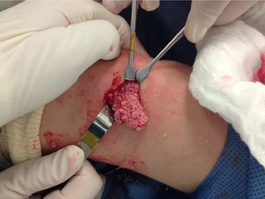
Macroscopic aspect of left knee synovium, showing moderate amount of whitish mass, similar to rice grains.
Discussion
Although imaging and laboratory studies suggest the diagnosis of JIA, synovial biopsy was requested in order to rule out sarcoidosis, not confirmed by histopathology. At surgery, a small amount of synovial fluid was found, along with the presence of a moderate amount of whitish mass that histopathology showed to be riziform bodies.
Riziform bodies are structures which can be found in synovial fluid or adhered to synovium, and have this denomination for its similar appearance to the rice grains. They consist of fibrin involving mononuclear cells, polymorphonuclear cells and red blood cells, and represent a nonspecific response to synovial inflammation.55 Martini G, Tregnaghi A, Bordin T, Visentin MT, Zulian F. Rice bodies imaging in juvenile idiopathic arthritis. J Rheumatol. 2003;30:2720-1.
6 DiVito A, Kan JH. Juvenile idiopathic arthritis with rice bodies. Pediatr Radiol. 2008;38:1263.
7 Chung C, Coley BD, Martin LC. Rice bodies in juvenile rheumatoid arthritis. AJR. 1998;170:698-700.-88 Forse CL, Mucha BL, Santos MLZ, Ongcapin EH. Rice body formation without rheumatic disease or tuberculosis infection: a case report and literature review. Clin Rheumatol. 2012;31:1753-6. They have already been described in several diseases such as tuberculous arthritis (where they were originally first reported in 1895), other infectious arthritis, osteoarthritis, rheumatoid arthritis and JIA.22 Huang G-S, Lee C-H, Chen C-Y. Clinical images: tuberculous rice bodies of the wrist. Arthritis Rheum. 2005;52:1950.
3 Bucki B, Lansaman J, Janson X, Billon-Galland MA, Marty C, Ruel M, et al. Osteoarthritis with rice bodies in calcium microcrystals 4 cases with ultrastructural study. Rev Rheum. 1994;61:415-20.
4 Jeong YM, Cho HY, Lee S-W, Hwang YM, Kim Y-K. Candida septic arthritis with rice body formation: a case report and review of literature. Korean J Radiol. 2013;14:465-9.
5 Martini G, Tregnaghi A, Bordin T, Visentin MT, Zulian F. Rice bodies imaging in juvenile idiopathic arthritis. J Rheumatol. 2003;30:2720-1.
6 DiVito A, Kan JH. Juvenile idiopathic arthritis with rice bodies. Pediatr Radiol. 2008;38:1263.
7 Chung C, Coley BD, Martin LC. Rice bodies in juvenile rheumatoid arthritis. AJR. 1998;170:698-700.
8 Forse CL, Mucha BL, Santos MLZ, Ongcapin EH. Rice body formation without rheumatic disease or tuberculosis infection: a case report and literature review. Clin Rheumatol. 2012;31:1753-6.
9 Druschel C, Funk JF, Kallinich T, Lieb A, Placzek RP. Development of rice bodies in 2 children younger than 3 years. J Clin Rheumatol. 2013;19:35-7.
10 Cox A, Allen R, Akikusa J. Rice bodies in juvenile idiopathic arthritis: a clinical image. J Paediatr Child Health. 2012;48:279-80.-1111 Teramoto A, Watanabe K, Kii Y, Kudo M, Otsubo H, Wada T, et al. Recurrent knee arthritis diagnosed as juvenile idiopathic arthritis with a 10-year asymptomatic period after arthroscopic synovectomy: a case report. J Med Case Rep. 2013;7:166-70.
Its etiopathogenesis is controversial. They can arise from areas of microinfarctions of inflamed synovium that are released into the SF and encapsulated by fibrin, abundantly produced in inflammatory processes of the synovium.77 Chung C, Coley BD, Martin LC. Rice bodies in juvenile rheumatoid arthritis. AJR. 1998;170:698-700.,99 Druschel C, Funk JF, Kallinich T, Lieb A, Placzek RP. Development of rice bodies in 2 children younger than 3 years. J Clin Rheumatol. 2013;19:35-7.,1010 Cox A, Allen R, Akikusa J. Rice bodies in juvenile idiopathic arthritis: a clinical image. J Paediatr Child Health. 2012;48:279-80.,1212 Wynne-Roberts CR, Cassidy JT. JRA with rice bodies: light and electric microscopic studies. Ann Rheum Dis. 1979;38:8-13. There is also another theory that suggests that riziform bodies are formed de novo in the SF, quite interesting for cases of osteoarthritis and the presence of apatite crystals and calcium pyrophosphate.44 Jeong YM, Cho HY, Lee S-W, Hwang YM, Kim Y-K. Candida septic arthritis with rice body formation: a case report and review of literature. Korean J Radiol. 2013;14:465-9.,88 Forse CL, Mucha BL, Santos MLZ, Ongcapin EH. Rice body formation without rheumatic disease or tuberculosis infection: a case report and literature review. Clin Rheumatol. 2012;31:1753-6. Rovenska et al. suggested that although there is a physiological lymphoangiogenesis associated with chronic inflammation in order to improve drainage of excessive SF, the formation of riziform bodies can be associated with the difficulty of lymphatic drainage of the inflamed synovial fluid.1313 Rovenska E, Stvrtina S, Greguska O, Pravda L, Rovensky J. Conspicuous synovial lymphatic capillaries in juvenile idiopathic arthritis synovitis with rice bodies. Ann Rheum Dis. 2005;64:328-9. The riziform bodies occur more often in knees and shoulders and we could say that they are the end products of inflammation, synovial proliferation and degeneration.99 Druschel C, Funk JF, Kallinich T, Lieb A, Placzek RP. Development of rice bodies in 2 children younger than 3 years. J Clin Rheumatol. 2013;19:35-7. On the other hand, there is a description of riziform bodies in the pleural fluid, tendon sheaths and bursae, suggesting a possibility of nonsynovial origin.22 Huang G-S, Lee C-H, Chen C-Y. Clinical images: tuberculous rice bodies of the wrist. Arthritis Rheum. 2005;52:1950.,88 Forse CL, Mucha BL, Santos MLZ, Ongcapin EH. Rice body formation without rheumatic disease or tuberculosis infection: a case report and literature review. Clin Rheumatol. 2012;31:1753-6.,1414 Chavan S, Sable SS, Tekade S, Punia P. Tuberculous tenosynovitis presenting as ganglion of wrist. Case Rep Surg. 2012;:143921.
The finding of riziform bodies in JIA was first reported by Wynne-Roberts et al. in 1979 in an 17-year-old adolescent1212 Wynne-Roberts CR, Cassidy JT. JRA with rice bodies: light and electric microscopic studies. Ann Rheum Dis. 1979;38:8-13. and, after, we found 7 more cases reported in the literature, with ages ranging from 2 to 17 years.55 Martini G, Tregnaghi A, Bordin T, Visentin MT, Zulian F. Rice bodies imaging in juvenile idiopathic arthritis. J Rheumatol. 2003;30:2720-1.
6 DiVito A, Kan JH. Juvenile idiopathic arthritis with rice bodies. Pediatr Radiol. 2008;38:1263.-77 Chung C, Coley BD, Martin LC. Rice bodies in juvenile rheumatoid arthritis. AJR. 1998;170:698-700.,99 Druschel C, Funk JF, Kallinich T, Lieb A, Placzek RP. Development of rice bodies in 2 children younger than 3 years. J Clin Rheumatol. 2013;19:35-7.
10 Cox A, Allen R, Akikusa J. Rice bodies in juvenile idiopathic arthritis: a clinical image. J Paediatr Child Health. 2012;48:279-80.-1111 Teramoto A, Watanabe K, Kii Y, Kudo M, Otsubo H, Wada T, et al. Recurrent knee arthritis diagnosed as juvenile idiopathic arthritis with a 10-year asymptomatic period after arthroscopic synovectomy: a case report. J Med Case Rep. 2013;7:166-70. This finding appears to be independent from arthritis severity and length of the disease.77 Chung C, Coley BD, Martin LC. Rice bodies in juvenile rheumatoid arthritis. AJR. 1998;170:698-700.,99 Druschel C, Funk JF, Kallinich T, Lieb A, Placzek RP. Development of rice bodies in 2 children younger than 3 years. J Clin Rheumatol. 2013;19:35-7.,1010 Cox A, Allen R, Akikusa J. Rice bodies in juvenile idiopathic arthritis: a clinical image. J Paediatr Child Health. 2012;48:279-80.,1515 Popert AJ, Scott DL, Wainwright AC, Walton KW, Williamson N, Chapman JH. Frequency of occurrence, mode of development, and significance of rice bodies in rheumatoid joints. Ann Rheum Dis. 1982;41:109-17. In the cases reported in children and adolescents, the time between the onset of arthritis and the finding of riziform bodies ranged from 2 months to 5 years, but not all reports contain this information. In our case, we observe that the appearance occurred after only 3 months of the onset of arthritis.
It would be interesting to conduct synovial biopsy to check for the presence of riziform bodies whenever the USG show the finding of debris. The difficulty of SF suction in a joint containing a large effusion may be due to the presence of riziform bodies and therefore the use of a thicker needle or a surgical approach may be necessary.1010 Cox A, Allen R, Akikusa J. Rice bodies in juvenile idiopathic arthritis: a clinical image. J Paediatr Child Health. 2012;48:279-80. The performance of synovial biopsy for checking the presence of riziform bodies is important, especially in cases of arthritis that is bulky and/or unresponsive to conventional treatment, as its withdrawal can cause symptomatic relief for the patient.77 Chung C, Coley BD, Martin LC. Rice bodies in juvenile rheumatoid arthritis. AJR. 1998;170:698-700.
Our objective was to describe what we believe is the 9th case reported about the presence of riziform bodies in JIA, and its presence should be more frequent than is reported in the literature and we are used to find.99 Druschel C, Funk JF, Kallinich T, Lieb A, Placzek RP. Development of rice bodies in 2 children younger than 3 years. J Clin Rheumatol. 2013;19:35-7. In rheumatoid arthritis, they can be found in 72% of joints, when searched.1515 Popert AJ, Scott DL, Wainwright AC, Walton KW, Williamson N, Chapman JH. Frequency of occurrence, mode of development, and significance of rice bodies in rheumatoid joints. Ann Rheum Dis. 1982;41:109-17. In general, we do not order synovial biopsy in other types of JIA other than the monoarticular and, with that, we are probably under-diagnosing the presence of riziform bodies, which may be responsible for large arthritis of difficult clinical treatment, but that respond well to surgical intervention.
-
☆
Work carried out in the Department of Rheumatology of the Center for Adolescent Health Studies, Universidade do Estado do Rio de Janeiro, Rio de Janeiro, RJ, Brazil.
References
-
1Oen KG, Cheang M. Epidemiology of chronic arthritis in childhood. Semin Arthritis Rheum. 1996;26:575-91.
-
2Huang G-S, Lee C-H, Chen C-Y. Clinical images: tuberculous rice bodies of the wrist. Arthritis Rheum. 2005;52:1950.
-
3Bucki B, Lansaman J, Janson X, Billon-Galland MA, Marty C, Ruel M, et al. Osteoarthritis with rice bodies in calcium microcrystals 4 cases with ultrastructural study. Rev Rheum. 1994;61:415-20.
-
4Jeong YM, Cho HY, Lee S-W, Hwang YM, Kim Y-K. Candida septic arthritis with rice body formation: a case report and review of literature. Korean J Radiol. 2013;14:465-9.
-
5Martini G, Tregnaghi A, Bordin T, Visentin MT, Zulian F. Rice bodies imaging in juvenile idiopathic arthritis. J Rheumatol. 2003;30:2720-1.
-
6DiVito A, Kan JH. Juvenile idiopathic arthritis with rice bodies. Pediatr Radiol. 2008;38:1263.
-
7Chung C, Coley BD, Martin LC. Rice bodies in juvenile rheumatoid arthritis. AJR. 1998;170:698-700.
-
8Forse CL, Mucha BL, Santos MLZ, Ongcapin EH. Rice body formation without rheumatic disease or tuberculosis infection: a case report and literature review. Clin Rheumatol. 2012;31:1753-6.
-
9Druschel C, Funk JF, Kallinich T, Lieb A, Placzek RP. Development of rice bodies in 2 children younger than 3 years. J Clin Rheumatol. 2013;19:35-7.
-
10Cox A, Allen R, Akikusa J. Rice bodies in juvenile idiopathic arthritis: a clinical image. J Paediatr Child Health. 2012;48:279-80.
-
11Teramoto A, Watanabe K, Kii Y, Kudo M, Otsubo H, Wada T, et al. Recurrent knee arthritis diagnosed as juvenile idiopathic arthritis with a 10-year asymptomatic period after arthroscopic synovectomy: a case report. J Med Case Rep. 2013;7:166-70.
-
12Wynne-Roberts CR, Cassidy JT. JRA with rice bodies: light and electric microscopic studies. Ann Rheum Dis. 1979;38:8-13.
-
13Rovenska E, Stvrtina S, Greguska O, Pravda L, Rovensky J. Conspicuous synovial lymphatic capillaries in juvenile idiopathic arthritis synovitis with rice bodies. Ann Rheum Dis. 2005;64:328-9.
-
14Chavan S, Sable SS, Tekade S, Punia P. Tuberculous tenosynovitis presenting as ganglion of wrist. Case Rep Surg. 2012;:143921.
-
15Popert AJ, Scott DL, Wainwright AC, Walton KW, Williamson N, Chapman JH. Frequency of occurrence, mode of development, and significance of rice bodies in rheumatoid joints. Ann Rheum Dis. 1982;41:109-17.
Publication Dates
-
Publication in this collection
Nov-Dec 2017
History
-
Received
17 Apr 2014 -
Accepted
14 Sept 2014