Abstract
Background
Computed tomography scans of the chest are often requested as a complementary examination to investigate a clinical suspicion of pulmonary disease caused by the novel coronavirus 19 (COVID-19).
Objectives
Our objective was to analyze the prevalence of incidental cardiovascular findings on chest CT scans requested to assess radiological signs suggestive of COVID-19 infection.
Methods
This cross-sectional, descriptive, and retrospective study reviewed 1,444 chest tomographies conducted in the Radiology department of the Hospital de Clínicas Gaspar Vianna, from March 1 to July 30, 2020, describing the prevalence of images suggestive of viral pneumonia by COVID-19 and incidental pulmonary and cardiovascular findings.
Results
The mean age of the patients was 50.6 ± 16.4 years and female sex was more frequent. Computed tomography without contrast was the most frequently used method (97.2%). Aortic and coronary wall calcification and cardiomegaly were the most prevalent cardiovascular findings. CT angiography revealed aortic aneurysms (9.7%), aortic dissection (7.3%) and thoracic aortic ulcers (2.4%).
Conclusions
Incidental cardiovascular findings occurred in about half of the chest CT scans of patients with suspected COVID-19, especially aortic calcifications, cardiomegaly, and coronary calcification.
Keywords:
incidental finding; cardiovascular finding; tomography; COVID-19
Resumo
Contexto
As tomografias de tórax são frequentemente solicitadas como exames complementares para avaliação de suspeita clínica de afecção pulmonar pelo novo coronavírus 19 (COVID-19).
Objetivos
Nosso objetivo foi analisar a prevalência dos achados cardiovasculares incidentais em tomografias de tórax solicitadas para avaliar sinais radiológicos sugestivos de COVID-19.
Métodos
Por meio de um estudo transversal, descritivo e retrospectivo, foram revisadas 1.444 tomografias de tórax realizadas no setor de radiologia do Hospital de Clínicas Gaspar Vianna, no período de 1° de março a 30 de julho de 2020, com a descrição da prevalência de imagens sugestivas de pneumonia viral pelo COVID-19, além de achados pulmonares e cardiovasculares incidentais.
Resultados
A média de idade dos pacientes foi 50,6±16,4 anos, sendo o sexo feminino o mais frequente. A tomografia sem contraste foi o método mais utilizado (97,2%), e opacidades em vidro fosco foram identificadas em 56,0% dos casos. Achados incidentais cardiovasculares ocorreram em 51,2% (intervalo de confiança 48,7%-53,8%) das tomografias, prevalecendo calcificação da parede aórtica (21,8%), cardiomegalia (10,5%), e calcificação coronária (5,0%). Nas tomografias com contraste, evidenciaram-se aneurismas de aorta (9,7%), dissecção de aorta (7,3%) e úlceras de aorta torácica (2,4%).
Conclusões
Achados cardiovasculares incidentais ocorreram em aproximadamente metade das tomografias de tórax de pacientes com suspeita de COVID-19, mais especificamente, calcificações da parede da aorta, cardiomegalia e calcificação coronária.
Palavras-chave:
achados incidentais; achado cardiovascular; tomografia; COVID-19
INTRODUCTION
Incidental findings in imaging exams are defined as unexpected findings unrelated to the scope of the clinical indication.11 Lumbreras B, Donat L, Hernandez-Aguado I. Incidental findings in imaging diagnostic tests: a systematic review. Br J Radiol. 2010;83(988):276-89. http://dx.doi.org/10.1259/bjr/98067945. PMid:20335439.
http://dx.doi.org/10.1259/bjr/98067945...
,22 Surov A, Bach AG, Schramm D. Clinically relevant cardiovascular findings detected on staging computed tomography in oncological patients. Angiology. 2016;67:630-7. http://dx.doi.org/10.1177/0003319715605971. PMid:26399716.
http://dx.doi.org/10.1177/00033197156059...
They are a common and well-known phenomenon in computed tomography (CT) examinations.33 Schramm D, Bach AG, Meyer HJ, Surov A. Thrombotic events as incidental finding on computed tomography in intensive care unit patients. Thromb Res. 2016;141:171-4. http://dx.doi.org/10.1016/j.thromres.2016.03.030. PMid:27058274.
http://dx.doi.org/10.1016/j.thromres.201...
However, the decisive factor is not the occurrence of a random finding, but its clinical relevance.11 Lumbreras B, Donat L, Hernandez-Aguado I. Incidental findings in imaging diagnostic tests: a systematic review. Br J Radiol. 2010;83(988):276-89. http://dx.doi.org/10.1259/bjr/98067945. PMid:20335439.
http://dx.doi.org/10.1259/bjr/98067945...
,33 Schramm D, Bach AG, Meyer HJ, Surov A. Thrombotic events as incidental finding on computed tomography in intensive care unit patients. Thromb Res. 2016;141:171-4. http://dx.doi.org/10.1016/j.thromres.2016.03.030. PMid:27058274.
http://dx.doi.org/10.1016/j.thromres.201...
In the literature on the subject, it is reported that incidental findings can be detected in up to 70% of all imaging investigations and, thankfully, the majority are of little clinical relevance.11 Lumbreras B, Donat L, Hernandez-Aguado I. Incidental findings in imaging diagnostic tests: a systematic review. Br J Radiol. 2010;83(988):276-89. http://dx.doi.org/10.1259/bjr/98067945. PMid:20335439.
http://dx.doi.org/10.1259/bjr/98067945...
2 Surov A, Bach AG, Schramm D. Clinically relevant cardiovascular findings detected on staging computed tomography in oncological patients. Angiology. 2016;67:630-7. http://dx.doi.org/10.1177/0003319715605971. PMid:26399716.
http://dx.doi.org/10.1177/00033197156059...
-33 Schramm D, Bach AG, Meyer HJ, Surov A. Thrombotic events as incidental finding on computed tomography in intensive care unit patients. Thromb Res. 2016;141:171-4. http://dx.doi.org/10.1016/j.thromres.2016.03.030. PMid:27058274.
http://dx.doi.org/10.1016/j.thromres.201...
These findings can be classified according to their clinical importance, as major, moderate, or minor.11 Lumbreras B, Donat L, Hernandez-Aguado I. Incidental findings in imaging diagnostic tests: a systematic review. Br J Radiol. 2010;83(988):276-89. http://dx.doi.org/10.1259/bjr/98067945. PMid:20335439.
http://dx.doi.org/10.1259/bjr/98067945...
From this perspective, newly-discovered tumors or aneurysms are of particular relevance, gallstones or pleural effusions can be considered moderately relevant, and uncomplicated liver cysts or simple vascular calcifications are irrelevant findings. The majority of incidental findings are clinically insignificant.11 Lumbreras B, Donat L, Hernandez-Aguado I. Incidental findings in imaging diagnostic tests: a systematic review. Br J Radiol. 2010;83(988):276-89. http://dx.doi.org/10.1259/bjr/98067945. PMid:20335439.
http://dx.doi.org/10.1259/bjr/98067945...
,22 Surov A, Bach AG, Schramm D. Clinically relevant cardiovascular findings detected on staging computed tomography in oncological patients. Angiology. 2016;67:630-7. http://dx.doi.org/10.1177/0003319715605971. PMid:26399716.
http://dx.doi.org/10.1177/00033197156059...
Cardiovascular incidental findings include disorders such as aneurysms, calcifications of cardiac valves and arteries, and thromboembolisms, which can sometimes be clinically asymptomatic and require further investigation, or may need therapeutic interventions, as is the case with giant aortic aneurysms.33 Schramm D, Bach AG, Meyer HJ, Surov A. Thrombotic events as incidental finding on computed tomography in intensive care unit patients. Thromb Res. 2016;141:171-4. http://dx.doi.org/10.1016/j.thromres.2016.03.030. PMid:27058274.
http://dx.doi.org/10.1016/j.thromres.201...
4 Jacobs PCA, Willem PTM, Diederick EG, van der Graaf Y. Prevalence of incidental findings in computed tomographic screening of the chest: a systematic review. J Comput Assist Tomogr. 2008;32(2):214-20. http://dx.doi.org/10.1097/RCT.0b013e3181585ff2. PMid:18379305.
http://dx.doi.org/10.1097/RCT.0b013e3181...
-55 Furtado CD, Aguirre DA, Sirlin CB, et al. Whole-body CT screening: spectrum of findings and recommendations in 1192 patients. Radiology. 2005;237(2):385-94. http://dx.doi.org/10.1148/radiol.2372041741. PMid:16170016.
http://dx.doi.org/10.1148/radiol.2372041...
Currently, because of the global health emergency faced by humanity and the scarcity of resources and limitations of large-scale confirmatory tests for the coronavirus 2019 disease (COVID-19), such as reverse transcription followed by polymerase chain reaction (RT-PCR),66 Kanne JP, Chest CT. Findings in 2019 Novel Coronavirus (2019- nCoV) Infections from Wuhan, China: Key Points for the Radiologist. Radiology. 2020;200241(1):16-7. http://dx.doi.org/10.1148/radiol.2020200241. PMid:32017662.
http://dx.doi.org/10.1148/radiol.2020200...
chest CT scans have come to play an important role supporting diagnosis because of their high sensitivity for detection of viral pneumonia and their potential to aid in assessment of disease progression and monitoring of treatment response.77 Orme NM, Fletcher JG, Siddiki HA, et al. Incidental findings in imaging research: evaluating incidence, benefit, and burden. Arch Intern Med. 2010;170(17):1525-32. http://dx.doi.org/10.1001/archinternmed.2010.317. PMid:20876402.
http://dx.doi.org/10.1001/archinternmed....
,88 Moyle P, Sonoda L, Britton P, Sinnatamby R. Incidental breast lesions detected on CT: what is their significance? Br J Radiol. 2010;83(987):233-40. http://dx.doi.org/10.1259/bjr/58729988. PMid:19546179.
http://dx.doi.org/10.1259/bjr/58729988...
As a result, it has become necessary to increasing use imaging techniques with satisfactory accuracy and ensure availability of tomographic images in the majority of emergency units, since this is the preferred diagnostic examination in cases of suspected viral infection by COVID-19. Another advantage of CT, in addition to investigations related to the clinical complaint, is its capacity to identify incidental abnormalities, i.e., findings unrelated to the indications for requesting it.66 Kanne JP, Chest CT. Findings in 2019 Novel Coronavirus (2019- nCoV) Infections from Wuhan, China: Key Points for the Radiologist. Radiology. 2020;200241(1):16-7. http://dx.doi.org/10.1148/radiol.2020200241. PMid:32017662.
http://dx.doi.org/10.1148/radiol.2020200...
7 Orme NM, Fletcher JG, Siddiki HA, et al. Incidental findings in imaging research: evaluating incidence, benefit, and burden. Arch Intern Med. 2010;170(17):1525-32. http://dx.doi.org/10.1001/archinternmed.2010.317. PMid:20876402.
http://dx.doi.org/10.1001/archinternmed....
8 Moyle P, Sonoda L, Britton P, Sinnatamby R. Incidental breast lesions detected on CT: what is their significance? Br J Radiol. 2010;83(987):233-40. http://dx.doi.org/10.1259/bjr/58729988. PMid:19546179.
http://dx.doi.org/10.1259/bjr/58729988...
9 Lee JH, Jeong SY, Kim YH. Clinical significance of incidental thyroid nodules identified on low-dose CT for lung cancer screening. Multidiscip Respir Med. 2013;8(1):56. http://dx.doi.org/10.1186/2049-6958-8-56. PMid:23985215.
http://dx.doi.org/10.1186/2049-6958-8-56...
-1010 Koos R, Kühl HP, Mühlenbruch G, Wildberger JE, Günther RW, Mahnken AH. Prevalence and clinical importance of aortic valve calcification detected incidentally on CT scans: comparison with echocardiography. Radiology. 2006;241(1):76-82. http://dx.doi.org/10.1148/radiol.2411051163. PMid:16908682.
http://dx.doi.org/10.1148/radiol.2411051...
Considering the above, in the present study we intend to analyze the prevalence of incidental cardiovascular findings in chest CTs performed because of suspected COVID-19 at a tertiary hospital in the state of Pará, Brazil.
METHOD
This is a retrospective and descriptive cross-sectional study conducted at the radiology service of the Hospital de Clínicas Gaspar Vianna (HCGV). The research corpus was collected at a general tertiary hospital specialized in cardiology, nephrology, and cardiovascular surgery, located in the city of Belém (PA).
This project was approved by the Research Ethics Committee at the institution and registered on the Plataforma Brasil (CAAE: 33706220.9.0000.0016) under ruling number 4.142.701.
Patients were identified using a computerized radiographic database that records all of the radiological studies performed by the radiology department. It was possible to include all chest CTs performed at the institution on patients from its various different sectors (wards, emergency department, or outpatients) from March 30 to July 2020 that had been requested because of indications related to respiratory symptoms, flu-like syndromes, or suspected COVID-19 pneumonia. CTs conducted because of other indications were excluded.
All CTs were performed using a Siemens Somatom Plus 16 multidetector scanner (Siemens Medical Systems Inc, Iselin, New Jersey, United States), and the tomographic images were recorded on a Vitria workstation and viewed with windows for bone (C800 W2000) and/or lung (C40 W80), both appropriate for the thorax. The examination was performed with the patient in dorsal decubitus with the following CT scan parameters: tube potential, 120 kVp; automatic tube current modulation, 30-70 mAs; pitch, 0.99-1.22 mm; matrix, 512 x 512; slice thickness, 1.0 mm; and field of view, 350 x 350 mm. Examinations reports were prepared by the institution’s three staff radiologists. When patients underwent more than one chest CT during the study period, only the results of the first examination were included in the analysis.
The research protocol comprised a list of 35 questions covering three principle topics: sociodemographic aspects, cardiovascular findings, and distribution of incidental findings.
Information was collected on the sample characteristics and organized in a spreadsheet in Microsoft® Office Excel® 2016.
The sample size was calculated based on an estimated prevalence of incidental cardiovascular findings of up to 60%, a standard error of 3.0%, and an alpha error of 5%, resulting in a minimum sample of 1,024 CT examinations.
Application of descriptive statistics involved drawing up tables and plotting graphs to present the results and calculating measures of position, such as arithmetic means and standard deviations.
The analytical statistics used to evaluate the results of variables for the sample were G tests and chi-square tests of fit for univariate tables. Descriptive and analytical statistics were computed using BioEstat® 5.4 software. A significance level of α = 0.05, or 5%, was adopted for decision making, indicating significant results with an asterisk (*).
RESULTS
During the period from March 1 through July 30, 2020, the HCGV radiology department performed a total of 1,444 chest CTs for suspected COVID-19 pulmonary infection.
Mean (standard deviation) patient age was 50.6±16.4 years. The youngest patient was 2 years old and the oldest was 99. There was a statistically significant difference (*p < 0.0001) between the proportions of age groups, with the greatest proportion of patients aged 40 to 49 years (25.7%).
There was a predominance of female patients (738; 51.1%), but the difference between sexes was not statistically significant (p = 0.4146).
The statistically significantly greatest proportion of requests for CT originated from the emergency department (48.8%), as shown in Table 1.
Epidemiological profile of patients who underwent computed tomography of the chest for suspected COVID-19 at the Hospital de Clínicas Gaspar Vianna, from March to July 2020.
The most common method used was CT without contrast (97.2%), used for a statistically significant proportion of the sample. Similarly, tomographic findings compatible with COVID-19 pulmonary involvement were observed in a significant 56% of the sample (*p = 0.0002) and the distribution pattern of pulmonary injury was < 25% ground-glass opacity in a statistically significant majority (*p < 0.0001) of patients, as shown in Table 2.
Data from Chest CTs for suspected COVID-19 at the Hospital de Clínicas Gaspar Vianna, from March to July 2020.
Analysis of the tomographic findings compatible with COVID-19 pulmonary involvement revealed that a statistically significant percentage of patients had ground-glass opacity (56%). Additional compatible findings included consolidation (18.4%), pleural effusion (12.6%), and parenchymal bands (7.5%); tomographic characteristics suggestive of COVID-19, among the other findings identified in the CT reports, as shown in Table 3.
Tomography findings compatible with COVID-19, from suspected COVID-19 cases at the Hospital de Clínicas Gaspar Vianna, March to July 2020.
Incidental pulmonary findings were reported in 63.2% of chest CTs. The finding with the greatest proportion was pulmonary nodule (18.3%), which was statistically the most significant (*p < 0.0001). The most frequent other findings were peribronchial thickening (13.1%), band atelectasis (11.4%), atelectasis (9.5%), and emphysema (5.7%), as shown in Table 4.
Incidental pulmonary findings on computed tomography of the chest in patients suspected of COVID-19, Hospital de Clínicas Gaspar Vianna, March to July 2020.
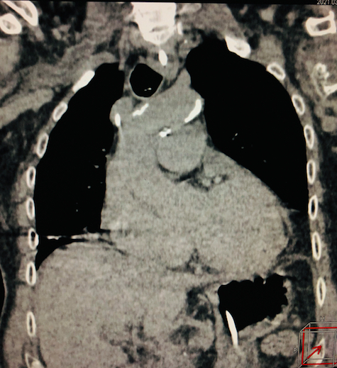
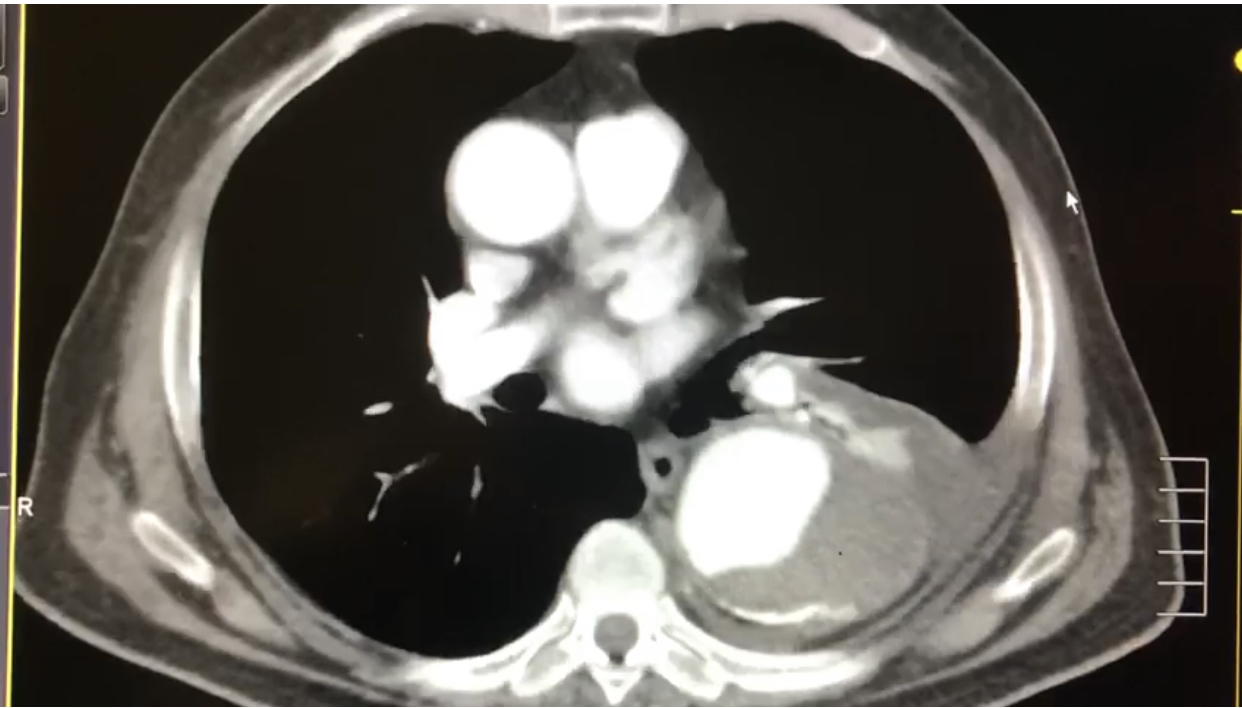
The database analysis was able to catalogue and distribute the cardiovascular findings present in 51.2% [confidence interval (CI) 48.7-53.8%] of the patients analyzed. The finding seen in the largest proportion of the sample was aortic wall calcification (21.8%), followed by cardiomegaly (10.5%), as illustrated in Table 5 and Figures 1 and 2. In turn, Table 6 shows the distribution of cardiovascular findings in the population over the age of 30 (1,299 patients). The most prevalent finding identified was aortic wall calcification (16.2%), followed by cardiomegaly (10.7%), and coronary calcification (4.5%).
Incidental cardiovascular findings on computed tomography of the chest in patients suspected of COVID-19, Hospital de Clínicas Gaspar Vianna, March to July 2020.

Incidental cardiovascular findings in computed tomography of the chest. Calcification of aortic and coronary wall.

Incidental cardiovascular findings in computed tomography of the chest. Aortic wall calcification and cardiomegaly.
Incidental cardiovascular findings on computed tomography of the chest in patients over the age of 30 suspected of COVID-19, Hospital de Clínicas Gaspar Vianna, March to July 2020.
With regard to aortic conditions, there were 12 incidental diagnoses of thoracic aorta aneurysm (0.83%), seven in the ascending aorta and five in the thoracic descending aorta. These 12 aneurysms were diagnosed in 10 patients (two male patients had two aneurysms each, in the ascending and descending aorta).
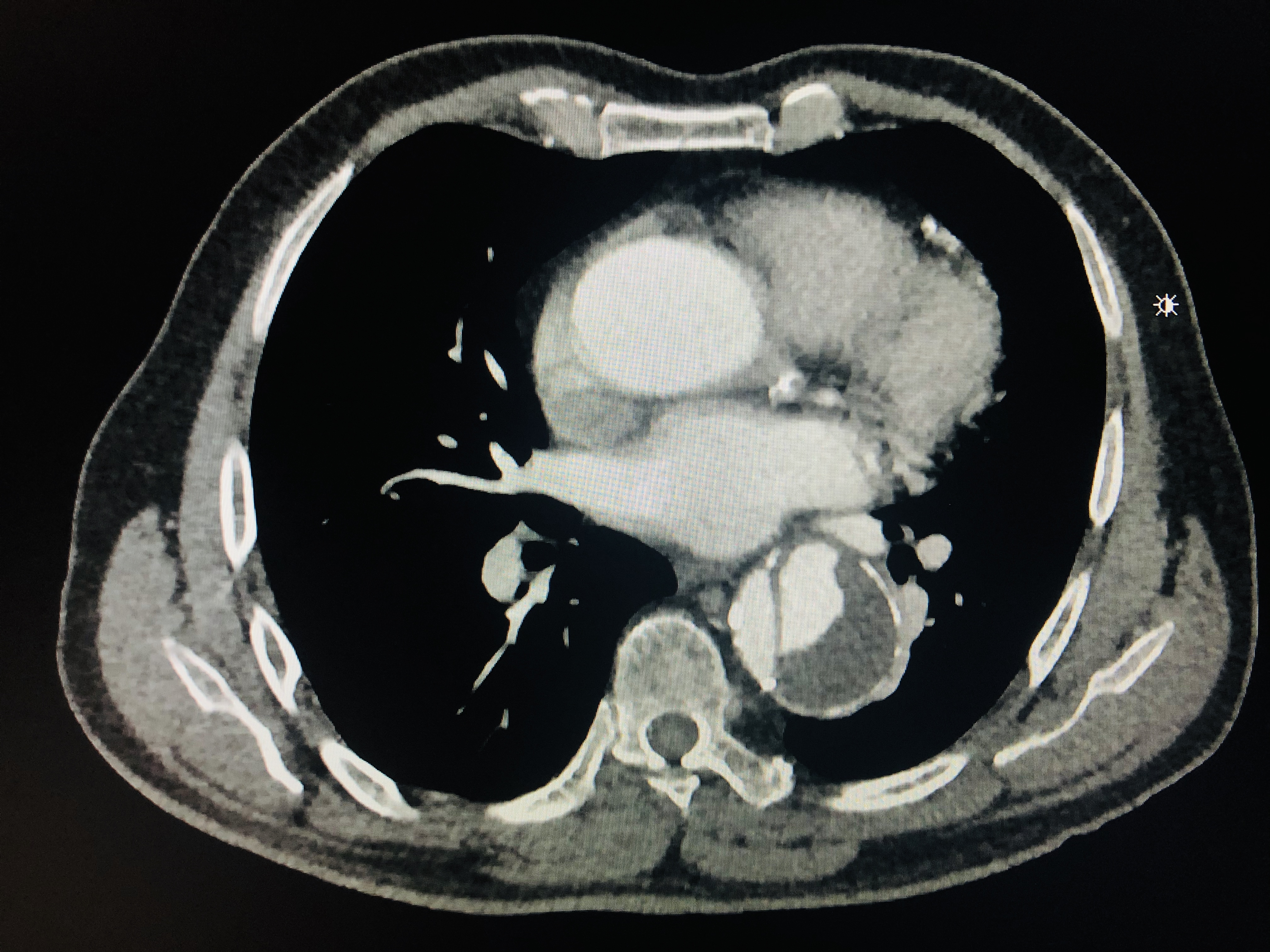
CT with contrast was conducted for 41 patients (2.8% of all scans), and yielded alternative diagnoses of aortic dissection in 7.3% (three of 41 scans) and thoracic aortic aneurysm in 9.7% (four of 41 scans). There were also findings of aortic ulcers in 2.4% (two of 41 scans). Figures 3 and 4 show examples of incidental aortic conditions detected with chest CT.

Incidental cardiovascular findings in computed tomography of the chest with contrast. Descending thoracic aortic aneurysm.

Incidental cardiovascular findings in computed tomography of the chest with contrast. Descending thoracic aorta dissection.
The prevalence of pulmonary thromboembolism (PTE) detected with computed tomography angiographies of the chest was 24.4% (10 of 41 examinations with contrast). Twenty-eight of these scans were of patients already in hospital and 13 were of patients referred from the emergency department. The incidental PTE findings were in two cases from the emergency department and eight of those already in hospital.
DISCUSSION
The important role played by diagnostic imaging has increased the number of CT examinations performed and, as CT became more widely used, radiologists began to observe a wave of findings unrelated to the clinical indications for which the examinations were ordered.11 Lumbreras B, Donat L, Hernandez-Aguado I. Incidental findings in imaging diagnostic tests: a systematic review. Br J Radiol. 2010;83(988):276-89. http://dx.doi.org/10.1259/bjr/98067945. PMid:20335439.
http://dx.doi.org/10.1259/bjr/98067945...
,33 Schramm D, Bach AG, Meyer HJ, Surov A. Thrombotic events as incidental finding on computed tomography in intensive care unit patients. Thromb Res. 2016;141:171-4. http://dx.doi.org/10.1016/j.thromres.2016.03.030. PMid:27058274.
http://dx.doi.org/10.1016/j.thromres.201...
,44 Jacobs PCA, Willem PTM, Diederick EG, van der Graaf Y. Prevalence of incidental findings in computed tomographic screening of the chest: a systematic review. J Comput Assist Tomogr. 2008;32(2):214-20. http://dx.doi.org/10.1097/RCT.0b013e3181585ff2. PMid:18379305.
http://dx.doi.org/10.1097/RCT.0b013e3181...
Confirmation of a COVID-19 diagnosis is based on RT-PCR of nasal or oropharyngeal samples collected with swabs. Patients infected by the SARS-Cov2 virus may present with changes seen on CT that are typical of the disease, such as ground-glass opacities. However, according to the Brazilian College of Radiology’s recommendations, CT should only be performed on symptomatic patients who have been admitted to hospital.1111 Bertolazzi P, Homero JF. A importância da Tomografia Computadorizada no diagnóstico da COVID-19. Arq Med. 2020;65(1):1-4. http://dx.doi.org/10.26432/1809-3019.2020.65.011.
http://dx.doi.org/10.26432/1809-3019.202...
Incidental findings are a well-known and common phenomenon in imaging exams.11 Lumbreras B, Donat L, Hernandez-Aguado I. Incidental findings in imaging diagnostic tests: a systematic review. Br J Radiol. 2010;83(988):276-89. http://dx.doi.org/10.1259/bjr/98067945. PMid:20335439.
http://dx.doi.org/10.1259/bjr/98067945...
,44 Jacobs PCA, Willem PTM, Diederick EG, van der Graaf Y. Prevalence of incidental findings in computed tomographic screening of the chest: a systematic review. J Comput Assist Tomogr. 2008;32(2):214-20. http://dx.doi.org/10.1097/RCT.0b013e3181585ff2. PMid:18379305.
http://dx.doi.org/10.1097/RCT.0b013e3181...
,77 Orme NM, Fletcher JG, Siddiki HA, et al. Incidental findings in imaging research: evaluating incidence, benefit, and burden. Arch Intern Med. 2010;170(17):1525-32. http://dx.doi.org/10.1001/archinternmed.2010.317. PMid:20876402.
http://dx.doi.org/10.1001/archinternmed....
,1212 Seo SG, Sung KH, Chung CY, et al. Incidental findings on knee radiographs in children and adolescents. Clin Orthop Surg. 2014;6(3):305-11. http://dx.doi.org/10.4055/cios.2014.6.3.305. PMid:25177456.
http://dx.doi.org/10.4055/cios.2014.6.3....
They can be classified according to their clinical importance, as major, moderate, or minor.1212 Seo SG, Sung KH, Chung CY, et al. Incidental findings on knee radiographs in children and adolescents. Clin Orthop Surg. 2014;6(3):305-11. http://dx.doi.org/10.4055/cios.2014.6.3.305. PMid:25177456.
http://dx.doi.org/10.4055/cios.2014.6.3....
Major findings include lesions suspected to be malignant diseases, such as thyroid nodules, changes to the thickness of the intestinal wall, and solid pancreatic or renal masses.1212 Seo SG, Sung KH, Chung CY, et al. Incidental findings on knee radiographs in children and adolescents. Clin Orthop Surg. 2014;6(3):305-11. http://dx.doi.org/10.4055/cios.2014.6.3.305. PMid:25177456.
http://dx.doi.org/10.4055/cios.2014.6.3....
Moderate findings, i.e. findings possibly of clinical relevance, include gallstones and pleural fluid accumulations or pleural effusion. Minor findings, or findings without clinical relevance, include simple renal or hepatic cysts, degenerative changes to the spinal column, and calcification of vessels. Many incidental findings are clinically insignificant.11 Lumbreras B, Donat L, Hernandez-Aguado I. Incidental findings in imaging diagnostic tests: a systematic review. Br J Radiol. 2010;83(988):276-89. http://dx.doi.org/10.1259/bjr/98067945. PMid:20335439.
http://dx.doi.org/10.1259/bjr/98067945...
,22 Surov A, Bach AG, Schramm D. Clinically relevant cardiovascular findings detected on staging computed tomography in oncological patients. Angiology. 2016;67:630-7. http://dx.doi.org/10.1177/0003319715605971. PMid:26399716.
http://dx.doi.org/10.1177/00033197156059...
,44 Jacobs PCA, Willem PTM, Diederick EG, van der Graaf Y. Prevalence of incidental findings in computed tomographic screening of the chest: a systematic review. J Comput Assist Tomogr. 2008;32(2):214-20. http://dx.doi.org/10.1097/RCT.0b013e3181585ff2. PMid:18379305.
http://dx.doi.org/10.1097/RCT.0b013e3181...
,77 Orme NM, Fletcher JG, Siddiki HA, et al. Incidental findings in imaging research: evaluating incidence, benefit, and burden. Arch Intern Med. 2010;170(17):1525-32. http://dx.doi.org/10.1001/archinternmed.2010.317. PMid:20876402.
http://dx.doi.org/10.1001/archinternmed....
This study found that female patients predominated and the most frequent source of patients was the institution’s emergency department, which requested 48.8% of the examinations.
The routine chest CT protocol used at the HCGV is a low-dose, 1.0 mm slice program without intravenous contrast. The great majority of chest CTs (97.2%) were performed without contrast, with contrast reserved for cases in which PTE or other vascular disorders requiring more detailed analysis were suspected.
The most common source of Chest CTs requests was the emergency department (48.8%). This elevated demand from external patients (rather than patients already admitted to the wards or intensive care unit) is probably because of the hospital’s characteristics, since it has an open-door emergency service for cardiovascular and nephrology patients.
Although CT is far from being the only examination of choice recommended for diagnosis by the various different medical specialty societies, it has become a valuable tool to support diagnosis in these patients, in addition to its utility for monitoring progress and detecting possible complications.1212 Seo SG, Sung KH, Chung CY, et al. Incidental findings on knee radiographs in children and adolescents. Clin Orthop Surg. 2014;6(3):305-11. http://dx.doi.org/10.4055/cios.2014.6.3.305. PMid:25177456.
http://dx.doi.org/10.4055/cios.2014.6.3....
,1313 Rosa ME, Matos MJ, Furtado RS, et al. Achados da COVID-19 identificados na tomografia computadorizada de tórax: ensaio pictórico. Einstein. 2020;18:eRW5741. http://dx.doi.org/10.31744/einstein_journal/2020RW5741.
http://dx.doi.org/10.31744/einstein_jour...
Thus, 56% of chest CTs requested had findings compatible with COVID-19 pulmonary involvement. The most characteristic findings were ground-glass opacity and consolidation; but many other findings can suggest pulmonary damage, depending on the disease phase and time since onset.1313 Rosa ME, Matos MJ, Furtado RS, et al. Achados da COVID-19 identificados na tomografia computadorizada de tórax: ensaio pictórico. Einstein. 2020;18:eRW5741. http://dx.doi.org/10.31744/einstein_journal/2020RW5741.
http://dx.doi.org/10.31744/einstein_jour...
A total of 63.2% of the CTs requested for diagnosis of presumed COVID-19 had incidental findings and the majority of these comprised pulmonary nodules (18.3%), peribronchial thickening (13.1%), band atelectasis (11.4%), and atelectasis (9.5%). Nodules were statistically significantly more common than the other pulmonary findings (p < 0.0001). It is believed that small nodules are not clinically relevant. However, eight patients exhibited findings of pulmonary masses larger than 3 cm (0.5% of the sample). This supports the hypothesis that the great majority of incidental findings are irrelevant, but the method can also detect asymptomatic injuries that need further management.1111 Bertolazzi P, Homero JF. A importância da Tomografia Computadorizada no diagnóstico da COVID-19. Arq Med. 2020;65(1):1-4. http://dx.doi.org/10.26432/1809-3019.2020.65.011.
http://dx.doi.org/10.26432/1809-3019.202...
Thus, although bilateral ground-glass opacity and consolidation are described as the predominant findings characteristic of imaging exams conducted for COVID-19, the manifestations seen on chest CT can vary from patient to patient and at different disease stages.1111 Bertolazzi P, Homero JF. A importância da Tomografia Computadorizada no diagnóstico da COVID-19. Arq Med. 2020;65(1):1-4. http://dx.doi.org/10.26432/1809-3019.2020.65.011.
http://dx.doi.org/10.26432/1809-3019.202...
Incidental cardiovascular findings were found in 51.2% (CI 48.7-53.8%) of the CT scans conducted and more than 40% had more than one finding. Calcification of aortic and coronary walls and cardiomegaly were the most common in the radiological reports. Less common findings involved hemorrhagic pericarditis and dilatation of the aortic root. These findings can be explained by the epidemiological profile observed in the study, since approximately 30% of the patients were over the age of 60, and also by the profile of the hospital’s patients in general, since it specializes in cardiovascular conditions and nephrology.
In an attempt to form more homogenous groups, a subset analysis was conducted after excluding data from 145 examinations of individuals less than 30 years old, leaving a subset of 1,299 CTs and revealing a similar distribution, with predominance of coronary and aorta wall calcification, cardiomegaly, and hemorrhagic pericarditis. Thus, regardless of its incidental nature early detection of potentially clinically relevant diseases, such as coronary calcification and aortic aneurysms or dissections, can change the prognosis of this population and have a positive impact on reduction of mortality and increase of quality of life.
Surov et al.22 Surov A, Bach AG, Schramm D. Clinically relevant cardiovascular findings detected on staging computed tomography in oncological patients. Angiology. 2016;67:630-7. http://dx.doi.org/10.1177/0003319715605971. PMid:26399716.
http://dx.doi.org/10.1177/00033197156059...
showed that cardiovascular findings can be identified in 6.8% of patients with malignant diseases investigated with CT. In contrast, Jacobs et al.44 Jacobs PCA, Willem PTM, Diederick EG, van der Graaf Y. Prevalence of incidental findings in computed tomographic screening of the chest: a systematic review. J Comput Assist Tomogr. 2008;32(2):214-20. http://dx.doi.org/10.1097/RCT.0b013e3181585ff2. PMid:18379305.
http://dx.doi.org/10.1097/RCT.0b013e3181...
reported that in their study the frequency of aortic aneurysm varied from 0.07% to 3.4%,44 Jacobs PCA, Willem PTM, Diederick EG, van der Graaf Y. Prevalence of incidental findings in computed tomographic screening of the chest: a systematic review. J Comput Assist Tomogr. 2008;32(2):214-20. http://dx.doi.org/10.1097/RCT.0b013e3181585ff2. PMid:18379305.
http://dx.doi.org/10.1097/RCT.0b013e3181...
while the frequency of aortic dissection varied from 0.06% to 0.2%.
Although incidental cardiac findings may not be relevant to the immediate clinical management of patients with suspected COVID-19 pulmonary involvement, they can influence long-term clinical management and improve prognosis. For example, coronary and aorta wall calcifications are known markers of atherosclerosis and signs of underlying cardiovascular disease, very often subclinical, which can influence primary prevention of atherosclerotic events.1414 Foley PWX, Hamaad A, El-Gendi H, Levya F. Incidental cardiac findings on computed tomography imaging of the thorax. BMC Res Notes. 2010;3(1):326. http://dx.doi.org/10.1186/1756-0500-3-326. PMid:21126380.
http://dx.doi.org/10.1186/1756-0500-3-32...
15 Takasu J, Budoff MJ, O’Brien KD, et al. Relationship between coronary artery and descending thoracic aortic calcification as detected by computed tomography: the Multi-Ethnic Study of Atherosclerosis. Atherosclerosis. 2009;204(2):440-6. http://dx.doi.org/10.1016/j.atherosclerosis.2008.09.041. PMid:19027115.
http://dx.doi.org/10.1016/j.atherosclero...
16 Ojha V, Mani A, Pandey NN, Sharma S, Kumar S. CT in coronavirus disease 2019 (COVID-19): a systematic review of chest CT findings in 4410 adult patients. Eur Radiol. 2020;30(11):6129-38. http://dx.doi.org/10.1007/s00330-020-06975-7. PMid:32474632.
http://dx.doi.org/10.1007/s00330-020-069...
-1717 Takasu J, Katz R, Nasir K, et al. Relationships of thoracic aortic wall calcification to cardiovascular risk factors: the Multi-Ethnic Study of Atherosclerosis (MESA). Am Heart J. 2008;155(4):765-71. http://dx.doi.org/10.1016/j.ahj.2007.11.019. PMid:18371491.
http://dx.doi.org/10.1016/j.ahj.2007.11....
On the other hand, cardiomegaly constitutes a late characteristic of left ventricular dysfunction and heart failure, both with poor prognosis, and could trigger further investigation and more effective cardiac treatment.1414 Foley PWX, Hamaad A, El-Gendi H, Levya F. Incidental cardiac findings on computed tomography imaging of the thorax. BMC Res Notes. 2010;3(1):326. http://dx.doi.org/10.1186/1756-0500-3-326. PMid:21126380.
http://dx.doi.org/10.1186/1756-0500-3-32...
Several different studies have shown that atherosclerotic plaques, calcifications, and aorta wall irregularities are very prevalent among patients with cardiovascular disease.1515 Takasu J, Budoff MJ, O’Brien KD, et al. Relationship between coronary artery and descending thoracic aortic calcification as detected by computed tomography: the Multi-Ethnic Study of Atherosclerosis. Atherosclerosis. 2009;204(2):440-6. http://dx.doi.org/10.1016/j.atherosclerosis.2008.09.041. PMid:19027115.
http://dx.doi.org/10.1016/j.atherosclero...
16 Ojha V, Mani A, Pandey NN, Sharma S, Kumar S. CT in coronavirus disease 2019 (COVID-19): a systematic review of chest CT findings in 4410 adult patients. Eur Radiol. 2020;30(11):6129-38. http://dx.doi.org/10.1007/s00330-020-06975-7. PMid:32474632.
http://dx.doi.org/10.1007/s00330-020-069...
-1717 Takasu J, Katz R, Nasir K, et al. Relationships of thoracic aortic wall calcification to cardiovascular risk factors: the Multi-Ethnic Study of Atherosclerosis (MESA). Am Heart J. 2008;155(4):765-71. http://dx.doi.org/10.1016/j.ahj.2007.11.019. PMid:18371491.
http://dx.doi.org/10.1016/j.ahj.2007.11....
Others have demonstrated that calcification of the descending aorta is related to calcification of the coronary arteries, an important predictor of cardiovascular disease. However, to date, no follow-up studies have been conducted to investigate the prognostic value of these abnormalities.1616 Ojha V, Mani A, Pandey NN, Sharma S, Kumar S. CT in coronavirus disease 2019 (COVID-19): a systematic review of chest CT findings in 4410 adult patients. Eur Radiol. 2020;30(11):6129-38. http://dx.doi.org/10.1007/s00330-020-06975-7. PMid:32474632.
http://dx.doi.org/10.1007/s00330-020-069...
,1717 Takasu J, Katz R, Nasir K, et al. Relationships of thoracic aortic wall calcification to cardiovascular risk factors: the Multi-Ethnic Study of Atherosclerosis (MESA). Am Heart J. 2008;155(4):765-71. http://dx.doi.org/10.1016/j.ahj.2007.11.019. PMid:18371491.
http://dx.doi.org/10.1016/j.ahj.2007.11....
Sverzellati et al.1818 Sverzellati N, Arcadi T, Salvolini L, et al. Under-reporting of cardio-vascular findings on chest CT. Radiol Med. 2016;121(3):190-9. http://dx.doi.org/10.1007/s11547-015-0595-0. PMid:26519045.
http://dx.doi.org/10.1007/s11547-015-059...
reported that 50% of 286 CT examinations ordered for pulmonary fibrosis, suspected pulmonary embolism, or lung cancer staging revealed potentially significant cardiovascular findings. Along the same lines, Choy et al.1919 Choy G, Kropil P, Scherer A, et al. Pertinent reportable incidental cardiac findings on chest CT without electrocardiography gating: review of 268 consecutive cases. Acta Radiol. 2013;54(4):396-400. http://dx.doi.org/10.1177/0284185113475918. PMid:23436832.
http://dx.doi.org/10.1177/02841851134759...
demonstrated that 61% of a consecutive series of routine chest CTs exhibited cardiac findings that merited reporting.
Previous studies of COVID-19 have shown that 63 to 67% of patients who died had cardiovascular comorbidities, most commonly hypertension, diabetes, and coronary cardiac disease,2020 Munden RF, Carter BW, Chiles C, et al. Managing Incidental Findings on Thoracic CT: Mediastinal and Cardiovascular Findings. A White Paper of the ACR Incidental Findings Committee. J Am Coll Radiol. 2018;15(8):1087-96. http://dx.doi.org/10.1016/j.jacr.2018.04.029. PMid:29941240.
http://dx.doi.org/10.1016/j.jacr.2018.04...
21 Zhou F, Yu T, Du R, et al. Clinical course and risk factors for mortality of adult inpa- tients with COVID-19 in Wuhan, China: a retrospective cohort study. Lancet. 2020;395(10229):1054-62. http://dx.doi.org/10.1016/S0140-6736(20)30566-3. PMid:32171076.
http://dx.doi.org/10.1016/S0140-6736(20)...
-2222 Chen T, Wu D, Chen H, et al. Clinical characteristics of 113 deceased patients with coronavirus disease 2019: retrospective study. BMJ. 2020;368:m1091. http://dx.doi.org/10.1136/bmj.m1091. PMid:32217556.
http://dx.doi.org/10.1136/bmj.m1091...
which are all factors that can be linked to vascular diseases.1919 Choy G, Kropil P, Scherer A, et al. Pertinent reportable incidental cardiac findings on chest CT without electrocardiography gating: review of 268 consecutive cases. Acta Radiol. 2013;54(4):396-400. http://dx.doi.org/10.1177/0284185113475918. PMid:23436832.
http://dx.doi.org/10.1177/02841851134759...
20 Munden RF, Carter BW, Chiles C, et al. Managing Incidental Findings on Thoracic CT: Mediastinal and Cardiovascular Findings. A White Paper of the ACR Incidental Findings Committee. J Am Coll Radiol. 2018;15(8):1087-96. http://dx.doi.org/10.1016/j.jacr.2018.04.029. PMid:29941240.
http://dx.doi.org/10.1016/j.jacr.2018.04...
21 Zhou F, Yu T, Du R, et al. Clinical course and risk factors for mortality of adult inpa- tients with COVID-19 in Wuhan, China: a retrospective cohort study. Lancet. 2020;395(10229):1054-62. http://dx.doi.org/10.1016/S0140-6736(20)30566-3. PMid:32171076.
http://dx.doi.org/10.1016/S0140-6736(20)...
-2222 Chen T, Wu D, Chen H, et al. Clinical characteristics of 113 deceased patients with coronavirus disease 2019: retrospective study. BMJ. 2020;368:m1091. http://dx.doi.org/10.1136/bmj.m1091. PMid:32217556.
http://dx.doi.org/10.1136/bmj.m1091...
Aortic conditions were detected incidentally on chest CT in up to 2.4% of cases, among which aneurysms were the most prevalent abnormality, especially in CTs without contrast. In the absence of intravenous contrast, significant aortic dissections and ulcerations are generally undetectable, which can be considered a limitation of the method.44 Jacobs PCA, Willem PTM, Diederick EG, van der Graaf Y. Prevalence of incidental findings in computed tomographic screening of the chest: a systematic review. J Comput Assist Tomogr. 2008;32(2):214-20. http://dx.doi.org/10.1097/RCT.0b013e3181585ff2. PMid:18379305.
http://dx.doi.org/10.1097/RCT.0b013e3181...
In the present study, just 2.8% of the CT scans were conducted with contrast. Aneurysms and dissections were observed in 9.7% and 7.3% respectively and suggested in the radiologist’s report in 1.2% of scans without intravenous contrast. On the other hand, PTE was observed in 24.4% of the patients who had scans with contrast, constituting a significant sample of cases admitted to the intensive care unit, where the more severe COVID-19 cases predominate.
Thus, is it is important to emphasize that incidental findings constitute an important event for patient clinical outcomes, since a considerable proportion of them have comorbidities concomitant to the novel coronavirus infection and, consequently, are more susceptible to the disease’s complications. If the incidental diagnosis is made early and is of a relevant nature, it increases the likelihood of management and prompt treatment and, consequently, of better prognosis for the infected population.
The present study is subject to limitations, since it is a retrospective study with selection bias, because it analyzed a sample of outpatients and inpatients from a specialist cardiovascular service and the data collection period was relatively short. The CT reports were prepared by three different radiologists and there was no analysis by race or other subsets. Some cardiovascular findings were observed in scans with contrast, including ulcers and dissections, which could indicate that CTs without contrast are limited in this respect.
No analysis was conducted of correlations between cardiovascular findings and risk factors because the study is based on CT scans and reports that were requested because of diagnosis suggestive of COVID-19. Additional studies are ongoing at the institution with research protocols that include better stratification of outpatients and inpatients in order to obtain more trustworthy and explanatory results.
CONCLUSIONS
Incidental cardiovascular findings were observed in approximately half of the chest CTs of patients with suspected COVID-19; more specifically, aortic calcifications, cardiomegaly, and coronary calcification.
-
How to cite: Reis JMC, Melo GS, Oliveira MV, et al. Incidental cardiovascular findings on chest CT scans requested for suspected COVID-19. J Vasc Bras. 2021;20:e20210052. https://doi.org/10.1590/1677-5449.210052
-
Financial support: None.
-
The study was carried out at Hospital de Clínicas Gaspar Vianna (HCGV), Belém, PA, Brazil.
REFERÊNCIAS
-
1Lumbreras B, Donat L, Hernandez-Aguado I. Incidental findings in imaging diagnostic tests: a systematic review. Br J Radiol. 2010;83(988):276-89. http://dx.doi.org/10.1259/bjr/98067945 PMid:20335439.
» http://dx.doi.org/10.1259/bjr/98067945 -
2Surov A, Bach AG, Schramm D. Clinically relevant cardiovascular findings detected on staging computed tomography in oncological patients. Angiology. 2016;67:630-7. http://dx.doi.org/10.1177/0003319715605971 PMid:26399716.
» http://dx.doi.org/10.1177/0003319715605971 -
3Schramm D, Bach AG, Meyer HJ, Surov A. Thrombotic events as incidental finding on computed tomography in intensive care unit patients. Thromb Res. 2016;141:171-4. http://dx.doi.org/10.1016/j.thromres.2016.03.030 PMid:27058274.
» http://dx.doi.org/10.1016/j.thromres.2016.03.030 -
4Jacobs PCA, Willem PTM, Diederick EG, van der Graaf Y. Prevalence of incidental findings in computed tomographic screening of the chest: a systematic review. J Comput Assist Tomogr. 2008;32(2):214-20. http://dx.doi.org/10.1097/RCT.0b013e3181585ff2 PMid:18379305.
» http://dx.doi.org/10.1097/RCT.0b013e3181585ff2 -
5Furtado CD, Aguirre DA, Sirlin CB, et al. Whole-body CT screening: spectrum of findings and recommendations in 1192 patients. Radiology. 2005;237(2):385-94. http://dx.doi.org/10.1148/radiol.2372041741 PMid:16170016.
» http://dx.doi.org/10.1148/radiol.2372041741 -
6Kanne JP, Chest CT. Findings in 2019 Novel Coronavirus (2019- nCoV) Infections from Wuhan, China: Key Points for the Radiologist. Radiology. 2020;200241(1):16-7. http://dx.doi.org/10.1148/radiol.2020200241 PMid:32017662.
» http://dx.doi.org/10.1148/radiol.2020200241 -
7Orme NM, Fletcher JG, Siddiki HA, et al. Incidental findings in imaging research: evaluating incidence, benefit, and burden. Arch Intern Med. 2010;170(17):1525-32. http://dx.doi.org/10.1001/archinternmed.2010.317 PMid:20876402.
» http://dx.doi.org/10.1001/archinternmed.2010.317 -
8Moyle P, Sonoda L, Britton P, Sinnatamby R. Incidental breast lesions detected on CT: what is their significance? Br J Radiol. 2010;83(987):233-40. http://dx.doi.org/10.1259/bjr/58729988 PMid:19546179.
» http://dx.doi.org/10.1259/bjr/58729988 -
9Lee JH, Jeong SY, Kim YH. Clinical significance of incidental thyroid nodules identified on low-dose CT for lung cancer screening. Multidiscip Respir Med. 2013;8(1):56. http://dx.doi.org/10.1186/2049-6958-8-56 PMid:23985215.
» http://dx.doi.org/10.1186/2049-6958-8-56 -
10Koos R, Kühl HP, Mühlenbruch G, Wildberger JE, Günther RW, Mahnken AH. Prevalence and clinical importance of aortic valve calcification detected incidentally on CT scans: comparison with echocardiography. Radiology. 2006;241(1):76-82. http://dx.doi.org/10.1148/radiol.2411051163 PMid:16908682.
» http://dx.doi.org/10.1148/radiol.2411051163 -
11Bertolazzi P, Homero JF. A importância da Tomografia Computadorizada no diagnóstico da COVID-19. Arq Med. 2020;65(1):1-4. http://dx.doi.org/10.26432/1809-3019.2020.65.011
» http://dx.doi.org/10.26432/1809-3019.2020.65.011 -
12Seo SG, Sung KH, Chung CY, et al. Incidental findings on knee radiographs in children and adolescents. Clin Orthop Surg. 2014;6(3):305-11. http://dx.doi.org/10.4055/cios.2014.6.3.305 PMid:25177456.
» http://dx.doi.org/10.4055/cios.2014.6.3.305 -
13Rosa ME, Matos MJ, Furtado RS, et al. Achados da COVID-19 identificados na tomografia computadorizada de tórax: ensaio pictórico. Einstein. 2020;18:eRW5741. http://dx.doi.org/10.31744/einstein_journal/2020RW5741
» http://dx.doi.org/10.31744/einstein_journal/2020RW5741 -
14Foley PWX, Hamaad A, El-Gendi H, Levya F. Incidental cardiac findings on computed tomography imaging of the thorax. BMC Res Notes. 2010;3(1):326. http://dx.doi.org/10.1186/1756-0500-3-326 PMid:21126380.
» http://dx.doi.org/10.1186/1756-0500-3-326 -
15Takasu J, Budoff MJ, O’Brien KD, et al. Relationship between coronary artery and descending thoracic aortic calcification as detected by computed tomography: the Multi-Ethnic Study of Atherosclerosis. Atherosclerosis. 2009;204(2):440-6. http://dx.doi.org/10.1016/j.atherosclerosis.2008.09.041 PMid:19027115.
» http://dx.doi.org/10.1016/j.atherosclerosis.2008.09.041 -
16Ojha V, Mani A, Pandey NN, Sharma S, Kumar S. CT in coronavirus disease 2019 (COVID-19): a systematic review of chest CT findings in 4410 adult patients. Eur Radiol. 2020;30(11):6129-38. http://dx.doi.org/10.1007/s00330-020-06975-7 PMid:32474632.
» http://dx.doi.org/10.1007/s00330-020-06975-7 -
17Takasu J, Katz R, Nasir K, et al. Relationships of thoracic aortic wall calcification to cardiovascular risk factors: the Multi-Ethnic Study of Atherosclerosis (MESA). Am Heart J. 2008;155(4):765-71. http://dx.doi.org/10.1016/j.ahj.2007.11.019 PMid:18371491.
» http://dx.doi.org/10.1016/j.ahj.2007.11.019 -
18Sverzellati N, Arcadi T, Salvolini L, et al. Under-reporting of cardio-vascular findings on chest CT. Radiol Med. 2016;121(3):190-9. http://dx.doi.org/10.1007/s11547-015-0595-0 PMid:26519045.
» http://dx.doi.org/10.1007/s11547-015-0595-0 -
19Choy G, Kropil P, Scherer A, et al. Pertinent reportable incidental cardiac findings on chest CT without electrocardiography gating: review of 268 consecutive cases. Acta Radiol. 2013;54(4):396-400. http://dx.doi.org/10.1177/0284185113475918 PMid:23436832.
» http://dx.doi.org/10.1177/0284185113475918 -
20Munden RF, Carter BW, Chiles C, et al. Managing Incidental Findings on Thoracic CT: Mediastinal and Cardiovascular Findings. A White Paper of the ACR Incidental Findings Committee. J Am Coll Radiol. 2018;15(8):1087-96. http://dx.doi.org/10.1016/j.jacr.2018.04.029 PMid:29941240.
» http://dx.doi.org/10.1016/j.jacr.2018.04.029 -
21Zhou F, Yu T, Du R, et al. Clinical course and risk factors for mortality of adult inpa- tients with COVID-19 in Wuhan, China: a retrospective cohort study. Lancet. 2020;395(10229):1054-62. http://dx.doi.org/10.1016/S0140-6736(20)30566-3 PMid:32171076.
» http://dx.doi.org/10.1016/S0140-6736(20)30566-3 -
22Chen T, Wu D, Chen H, et al. Clinical characteristics of 113 deceased patients with coronavirus disease 2019: retrospective study. BMJ. 2020;368:m1091. http://dx.doi.org/10.1136/bmj.m1091 PMid:32217556.
» http://dx.doi.org/10.1136/bmj.m1091
Publication Dates
-
Publication in this collection
07 Jan 2022 -
Date of issue
2021
History
-
Received
02 Apr 2021 -
Accepted
07 Oct 2021