Abstracts
PURPOSE: To study the integration of the external layer of the calvaria's frontal bone to repair craniofacial skeleton injuries in primates. METHODS: Ten Rhesus monkeys underwent surgery in two stages. In the first stage, four bone fragments were harvested from the external layer of the calvaria's frontal bone and were transplanted two on the calvaria's frontal bone and the other two onto the maxillary bone, by onlay and inlay. Seven weeks thereafter, four fragments were extracted from the transplantated area. The animals were not sacrificed. RESULTS: Macroscopic examination reveals bone regeneration, the areas onto which the bone fragments were transplantated having consequently increased in volume. The results of optical and electron microscopy is being carried out.
Skull; Bone transplantation; Macaca mulatta
OBJETIVO: Estudar a integração da lâmina externa da calvária, na reparação de lesões do esqueleto craniofacial em primatas. MÉTODOS: Foram operados dez macacos Rhesus em duas etapas. Na primeira foram retirados quatro fragmentos ósseos da lâmina externa da região frontal da calvária e transplantados, dois na calvária e dois no osso maxilar por aposição e reposição. Sete semanas após, foram retirados quatro fragmentos dos locais transplantados. Os animais não foram sacrificados. RESULTADOS: Macroscopicamente evidenciou-se regeneração óssea, com conseqüente aumento de volume dos leitos receptores na face. Aguarda-se análise da microscopia óptica e eletrônica.
Crânio; Transplante ósseo; Macaca mulatta
2 PRELIMINARY NOTE
The use of the external layer of the calvaria's frontal bone to repair craniofacial skeleton injuries in Macaca mulatta (Rhesus)1 1 Work performed in the Departement of Plastic Surgery of the Medical School Fundação ABC
Emprego da lâmina externa da região frontal da calvária na reparação de lesões do esqueleto craniofacial em Macaca mulatta (Rhesus)
José Mário Camelo-NunesI, 2 2 Professor of Plastic Surgery of the Medical School of the Fundação ABC, São Paulo, Brazil ; Ubimara Pereira RodriguesII,3 3 Technical Director of the Central Biotic Resources Division of the Instituto Butantan, São Paulo, Brazil ; Saul GoldenbergIII
IProfessor of Plastic Surgery of the Medical School of the Fundação ABC, São Paulo, Brazil
IITechnical Director of the Central Biotic Resources Division of the Instituto Butantan, São Paulo, Brazil
IIIChairman Department of Surgery Federal University of São Paulo, Brazil
Address to correspondence Address to correspondence José Mário Camelo-Nunes R. Borges Lagoa, 783/12 04038-031 São Paulo, SP Tel.: (11)5579-8938 camelonunes@aol.com
ABSTRACT
PURPOSE: To study the integration of the external layer of the calvaria's frontal bone to repair craniofacial skeleton injuries in primates.
METHODS: Ten Rhesus monkeys underwent surgery in two stages. In the first stage, four bone fragments were harvested from the external layer of the calvaria's frontal bone and were transplanted two on the calvaria's frontal bone and the other two onto the maxillary bone, by onlay and inlay. Seven weeks thereafter, four fragments were extracted from the transplantated area. The animals were not sacrificed.
RESULTS: Macroscopic examination reveals bone regeneration, the areas onto which the bone fragments were transplantated having consequently increased in volume. The results of optical and electron microscopy is being carried out.
Keywords: Skull. Bone transplantation. Macaca mulatta.
RESUMO
OBJETIVO: Estudar a integração da lâmina externa da calvária, na reparação de lesões do esqueleto craniofacial em primatas.
MÉTODOS: Foram operados dez macacos Rhesus em duas etapas. Na primeira foram retirados quatro fragmentos ósseos da lâmina externa da região frontal da calvária e transplantados, dois na calvária e dois no osso maxilar por aposição e reposição. Sete semanas após, foram retirados quatro fragmentos dos locais transplantados. Os animais não foram sacrificados.
RESULTADOS: Macroscopicamente evidenciou-se regeneração óssea, com conseqüente aumento de volume dos leitos receptores na face. Aguarda-se análise da microscopia óptica e eletrônica.
Descritores: Crânio. Transplante ósseo. Macaca mulatta.
Introduction
The use of the calvaria's frontal bone as a source of bone donating tissue for the treatment of craniofacial skeleton deformities (congenital and acquired) was popularized in 1982, by Tessier (1).
In humans, these transplants are generally harvested from the parietal bone, specifically from its external layer, which allows the cranial cavity to remain intact (2 - 6).
In our review of the pertinent literature found no research on the use of the external layer of the calvaria's frontal bone to repair craniofacial skeleton injuries of non-human primates.
Methods
Sample
Consisted of ten mammals of the Primate order, Catarrhini infraorder (Old World monkeys), Cercophitecos family, Macaca genus, and Mulatta species (Rhesus).
In January 1999 the birth of ten animals was programmed. These were specifically used for this study.
The sample consisted on seven male monkeys and three female. The animals weight were between 1992,0 g to 3128,0 g and 19 to 23 months old.
The experimental protocol was approved by the Animal Research Ethics Comittee of the Medical School of the Fundação ABC, São Paulo - Brazil.
The monkeys were severely controlled with regard to both ecto- and endo-parasites, tuberculinization, dwelling, physical and chemical restraint, feeding, water supply, fluid extraction, mating, and control of growth and development (7).
Procedures
The animals were submitted to pre-surgical tests (hemogram, coagulation profile and glycemia), were considered healthy and made available for the surgical procedures. All underwent a two-hour pre-surgical fast.
Simple x-rays of the head, fontal and profile positions, and of the closed maxilla and mandible were taken and moulds of the dental arcade were cast in order to produce gypsum models. All stages of research (including pre- and post-surgery) were photographed, both conventionally and digitally, and filmed.
The animals were given 0.3mg/Kg of a cetamine hydrochloride solution and 7.5mg/Kg of 2-(2.6 xilidine)-5,6-dihydro-4H-1,3-tiazine intramuscularly as pre-anaesthesic medication.
They were considered to be pre-anaesthetized on average after four minutes, by which time they had lost their aggressiveness, were in a lateral decubitus position, open-eyed and with miotic pupils.
Anaesthesia was controlled by means of pulmonary and cardiac auscultation, peripheral blood oxygen saturation, peripheral pulse count and observation of pupil reflexes and eyelid movement.
Anaesthesia was maintained with cetamine hydrochloride, an average of 2.25 doses of 16.75mg/Kg each having been used at average intervals of 58.25 minutes.
Local anaesthesia of the scalp and of the oral cavity was applied through the infiltration of a solution with bupivacaine hydrochloride (5mg/ml), lidocain hydrochloride (20mg/ml) and epinephrine with a final concentration of 1:400,000.
The monkeys were placed on the surgical table in a horizontal dorsal decubitus position, the head partially flexed with the help of a pad. The upper limbs were maintained in an abducted position, by bandaging and the lower limbs were extended and immobilized in the same way; the sensor for peripheral monitoring was attached to the left foot.
The surgical team consisted of one surgeon, one anaesthetist, two assistants and one surgical technologist handling the instrumentation.
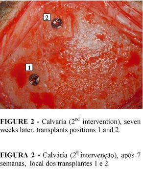
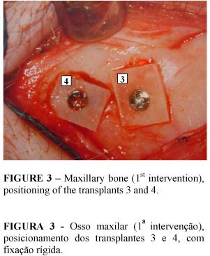
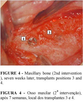
The study was divided into two surgical stages. In the first intervention four calvaria's bone fragments were harvested from the external layer of the calvaria's frontal bone. Two of them were transplantated onto the calvaria's frontal bone (one on the same donating place onlay and the other on the calvaria's frontal bone inlay) and the other two onto the maxilla (one defected by means of bone wear away onlay and the other on the maxilla inlay) and fixed with titanium microscrews (Osteomed TM).
Seven weeks later, four fragments were removed from the aforementioned transplants. All the fragments were measured with digital calipers accurate to the thousandth of a millimeter as regards their vertical and horizontal dimensions, as well as their thickness.
Preliminary Results
Preliminary macroscopic results show bone regeneration with the volume of the facial areas onto which the bone fragments were grafted having consequently increased (Figures 1, 2, 3 and 4).
Microscopic study involving optical microscopy and electron micrography is currently being carried out.
No complications arose during the post-surgical period and all the animals were monitored on a daily basis and analyzed, under anaesthesia, 21 days after each of the two interventions. The animals are currently back in their natural habitat.
The data and illustrations are in the process of being recorded in the respective protocols for subsequent publishing.
Acknowledgements
Andressa M. C. Inai, Carlos A. Andreucci, Cassio M. R. Amaral, Dirceu Solé, Eduardo F. Simões, Elie Fiss, Elizabeth J. G. Valentini, Ercole S. Neto, Eric Solyom, Fernão S. Oliveira, Gerson V. P. Filho, Giancarlo Dall'Olio, Hisakazu Hayashi, Jean-François Tulasne, Juliana B. Garcia, Ligia M. C. Nunes, Neil F. Novo, Osvaldo Mora, Paul Tessier, Paulo O. Gomes, Renato Albieri, Saul Goldenberg, Teresa R. B. Adão, Vânia G. M. Mattaraia.
Data do recebimento: 10/12/2002
Data da revisão: 14/01/2003
Data da aprovação: 04/02/2003
Conflito de interesse: nenhum
Fonte de financiamento: nenhuma
- 1. Tessier P. Autogenous bone grafts taken from the calvarium for facial and cranial applications. Clin Plast Surg 1982; 9: 531-8.
- 2. Kline RM, Wolfe SA. Complications associated with the harvesting of cranial bone grafts. Plast Reconstr Surg 1995; 95: 5-20.
- 3. Tulasne JF, Camelo-Nunez JM. Greffe osseuse crânienne intra-sinusienne. J de parondontologie & d'implantologie orale 1999; 18: 183-97.
- 4. Camelo-Nunes JM, Pereira Filho GV, Spada Neto E, Dall'Olio G. Enxerto ósseo craniano intra-sinusiano. Arq Med ABC 2001; 24: 60-9.
- 5. Ishizuka MMA. Contribuição ao estudo da espessura da calota craniana [thesis]. São Paulo: Universidade Federal de São Paulo; 1987.
- 6. Gomes Filho, WR. Espessura do enxerto de calota craniana e crista ilíaca alveolar da maxila como base nos implantes em titânio [thesis]. São Paulo: Universidade Federal de São Paulo; 2001.
-
7Sanitary control program of the primate (rhesus) colony of the Instituto Butantan, São Paulo, 2002.
Publication Dates
-
Publication in this collection
10 Nov 2004 -
Date of issue
Mar 2003
History
-
Accepted
04 Feb 2003 -
Reviewed
14 Jan 2003 -
Received
10 Dec 2002