Abstract
Suid herpesvirus 1 (SuHV-1) is the causative agent of pseudorabies (PR), a disease of great importance due to the huge losses it causes in the swine industry. The aim of this study was to determine a method for genotyping SuHV-1 based on partial sequences of the gene coding for glycoprotein C (gC) and to elucidate the possible reasons for the variability of this region. A total of 109 gCsequences collected from GenBank were divided into five major groups after reconstruction of a phylogenetic tree by Bayesian inference. The analysis showed that a portion of gC (approximately 671 bp) is under selective pressure at various points that coincide with regions of protein disorder. It was also possible to divide SuHV-1 into five genotypes that evolved under different selective pressures. These genotypes are not specific to countries or continents, perhaps due to multiple introduction events related to the importation of swine.
pseudorabies; glycoprotein; bioinformatics
GENETICS AND MOLECULAR MICROBIOLOGY
Pseudorabies virus can be classified into five genotypes using partial sequences of UL44
Fonseca Jr., A. A.I* * Corresponding Author. Mailing address: Laboratório Nacional Agropecuário, MAPA, Pedro Leopoldo-MG.; E-mail: antonio.a.fonsecajr@gmail.com ; Camargos, M. F.I; Sales, M. L.I; Heinemann, M. B.II; Leite, R.C.II; Reis, J. K. P.II
ILaboratório Nacional Agropecuário, Ministério da Agricultura, Pecuária e Abastecimento, Pedro Leopoldo, MG, Brasil
IILaboratório de Retroviroses, Escola de Veterinária, Universidade Federal de Minas Gerais, Belo Horizonte, MG, Brasil
ABSTRACT
Suid herpesvirus 1 (SuHV-1) is the causative agent of pseudorabies (PR), a disease of great importance due to the huge losses it causes in the swine industry. The aim of this study was to determine a method for genotyping SuHV-1 based on partial sequences of the gene coding for glycoprotein C (gC) and to elucidate the possible reasons for the variability of this region. A total of 109 gCsequences collected from GenBank were divided into five major groups after reconstruction of a phylogenetic tree by Bayesian inference. The analysis showed that a portion of gC (approximately 671 bp) is under selective pressure at various points that coincide with regions of protein disorder. It was also possible to divide SuHV-1 into five genotypes that evolved under different selective pressures. These genotypes are not specific to countries or continents, perhaps due to multiple introduction events related to the importation of swine.
Key words: pseudorabies, glycoprotein, bioinformatics
INTRODUCTION
Suid herpesvirus 1 (SuHV-1) is the causative agent of pseudorabies (PR), a disease with major importance in swine industry because it causes considerable losses in the production chain. PR is listed as one of the most important diseases affecting pigs in the Manual of Diagnostic Tests and Vaccines for Terrestrial Animals of the World Organization for Animal Health (34). SuHV-1 is a member of the Herpesviridae family, subfamily Alphaherpesvirinae, and has only one serotype. The natural host of this virus is the pig; however, it can infect other domestic animals such as dogs and cattle, causing encephalitis with almost 100% lethality (19).
SuHV-1 is classified into different genotypes based on restriction endonuclease analysis (3), but more recent studies conducted in Brazil, the United States and Europe have characterized viral strains based on partial sequencing of UL44 focusing on the region encoding the N-terminal portion of glycoprotein C (10,9, 20, 12). The UL44 gene is one of the most variable regions of the SuHV-1 genome (17). The gene encodes glycoprotein C (gC), which is the main component involved in adhesion to host cell receptors and is considered to be a potent inducer of the immune response. The protein contains eight N-glycosylation sites and three redundant heparin-binding domains (HBDs) (32). This glycoprotein is involved in two distinct steps of virus adhesion to host cells. The first step is an initial, low-affinity interaction between gC and cellular heparin-like receptors, followed by a second interaction that results in a more stable binding of the virus to the cells (36).
The use of molecular data such as nucleotide and amino acid sequences is an essential tool for understanding the variability and epidemiology of the virus. These tools were used to analyze outbreaks occurring in areas of high swine production in Brazil between 1983 and 2003 (9) and in the United States in 1989 (10). Another relevant analysis is the genetic profiling of the strains circulating in wild boars or pigs (10, 12) or the investigation of the spread of live vaccine strains among feral pigs (12). These surveys found a high degree of conservation between virus sequences and did not indicate a correlation between outbreak location and phylogenetic groups for SuHV-1. Another common feature in this field is that most research is restricted to the study of SuHV-1 genetic variation in specific territories and does not involve other methods of tree reconstruction besides neighborjoining.
Other methods such as maximum likelihood (ML) and Bayesian inference (BI) have been used for phylogenetic analysis based on amino acids and nucleotide substitutions. These methods have been used in several studies, including molecular biology analyses of viruses from various countries such as genotyping of bovine leukemia virus, and have produced useful and interesting new data (26). These bioinformatic tools can also be used to study polymorphism and selective pressure in SuHV-1 nucleotide sequences.
The aim of this study was to use new bioinformatics tools such as ML and BI for analysis of partial sequences of the UL44 gene available in the GenBank. This analysis will lead to a better understanding of the relationship between SuHV-1 isolates from different regions of the globe and the selective pressure and polymorphism found in each group.
MATERIALS AND METHODS
SuHV Sequences
We obtained complete or partial nucleotide sequences of SuHVUL44 from GenBank. All UL44 sequences were analyzed in previous studies (10, 9, 20, 12), with the exception of sequences from China, South Korea and Malaysia. The 109 sequences were named according to the name of the isolate in GenBank, followed by a three letter code to identify the country: Germany (GER), Brazil (BRA), China (CHI), North Korea (SKO), Slovakia (SLK), Spain (ESP), United States (USA), Japan (JAP), Hungary (HUN), Northern Ireland (NIR) and Malaysia (MAL). In addition, three sequences are present for the Bartha vaccine: one from a complete genome sequence, one from Brazil and another from China.
In addition, we sequenced 12 SuHV-1 samples from outbreaks occurring between 2002 and 2003 in the state of Santa Catarina in Brazil in accordance with Goldberg et al. (10).
SuHV-1 Phylogenetic reconstruction
Nucleotide sequences were submitted to three programs for the reconstruction of phylogenetic trees. The MEGA 4.0 program (18) was used to reconstruct a phylogeny by neighbor joining (using the maximum composite likelihood model) with 1000 bootstrap replicates (31). The best model for the reconstruction of phylogenetic trees was selected using the jModelTest program. The chosen model was the TnR93 with gamma distribution and optimized frequencies of substitution (24).
After choosing the model, reconstruction of the phylogeny by the method of maximum likelihood was performed using the Seaview program (11). The parameters generated by the jModelTest program were also included in the MrBayes program to create trees using the Bayesian method (16). Additionally, using MEGA 4.0, the mean distances between and within the groups formed in the phylogenetic trees were determined based on the number of differences.
Evolutionary history of Brazilian SuHV-1 isolates
In addition to the reconstruction of phylogenetic trees as described above, another type of analysis was performed. The Brazilian samples were chosen to trace the history of the entry of SuHV-1 into the country. The Brazilian samples were selected because of the greater knowledge of our group regarding the isolates and the history of PR in Brazil compared to other nations. Brazilian sequences and sequences from other countries with dates of isolation from each group identified previously were submitted to the program Beast (5) with substitution models defined by jModelTest. Codons were grouped into three partitions, and the substitution model was unlinked across codon positions. Each analysis was run such that the effective sample size was greater than 200. Samples from group E were not included because we could not find any information about the date of isolation of the virus sequenced.
Predictive analysis of translated sequences
All analyses of the translated sequences were generated using strain NIA-3 as a reference (GenBank accession number D49437). Sequences for the Shope and Bartha strains were also included in this analysis.
Sequences from the five groups were uploaded to the ELM Functional Sites in Proteins website (available at http://elm.eu.org/) for analysis and confirmation of glycosylation domains. Analysis of hydrophobicity and hydrophilicity was performed using the Kyte and Doolittle mean hydrophobicity method (seven-residue scan window without gaps) and the Hopp and Woodsmean hydrophilicity method (six-residue scan window without gaps) in the BioEdit program. The analysis focused on the signal peptide, formed by the first 22 amino acids, six glycosylation sites and three heparin-binding domains.
The SIFT program (available at http://blocks.fhcrc.org/sift/SIFT.html) (21) was used to predict the degree of tolerance of amino acid substitutions. Analysis of the influence of amino acid substitutions on the secondary structure and solvent accessibility was performed using the nnPredict (available at http://alexander.compbio.ucsf.edu/~nomi/nnpredict.html) and Scratch (available at http://www.ics.uci.edu/~baldig/scratch/index.html) programs. The domain search was performed using the ProDom program (available at http://prodom.prabi.fr/prodom/current/html/home.php).
Identification of specific positive and negative selection sites
The SuHV-1 UL44 sequences were submitted to search for specific positive and negative selection sites performed by the Selecton program (30; available at http://selecton.bioinfo.tau.ac.il/). The chosen model was M8, the default model for all Selecton runs. This model allows for positive selection operating on the protein. The results were statistically verified with the null model M8a, which is similar to the M8 model, except for not allowing for positive selection. Thus, only neutral and purifying selections are allowed. This null model allows for testing the hypothesis by performing a likelihood ratio test between the two models to see which model fits the data better.
All sequences were tested using a single file. Afterward, each group found in the phylogenetic tree was tested separated to verify its involvement under different selective pressures.
RESULTS
Phylogenetic tree reconstruction
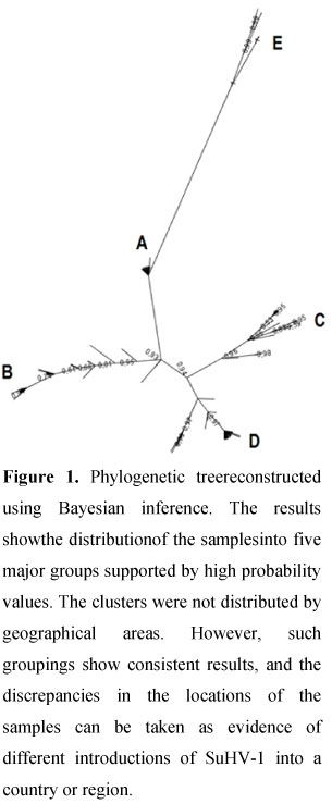
The results of the phylogenetic analysis showed several differences, especially among more distantly related samples (Figure 1). Some eastern samples were placed more distantly using NJ but were positioned next to a group of European isolates using ML and BI. Another difference in phylogeny was the peculiar position of IB341BRA, which was similar using NJ and BI but different using ML. The three sequences for the Bartha vaccine were positioned in different groups. BarthaBRA and BarthaPLOS were positioned in a group of European sequences, and BarthaCHI grouped with samples from China.
Analyses using ML and BI formed five major groups supported by high bootstrap values and credibility (Fig. 1). These groups were not as clear using NJ. The groups were named A, B, C, D and E and were characterized primarily by a hotspot between residues 184 and 195 (Fig. 2). The sequences originating from viruses isolated in Europe were distributed among all of the groups.
The distances calculated within each group were 0.1 for A, 3.7 for B, 3.6 for C, 2.7 for D and 7.6 for E. All of these distances were smaller than the distances calculated between groups, as can be seen in Table 1.
Evolutionary history of Brazilian SuHV-1 isolates
The results of the analysis of the Brazilian samples allowed us to estimate the divergence time (in years) relative to other countries. The time of divergence was 264 years for groups C and D and 410 years for groups C and B. It was not possible to calculate a clear time of divergence between group A and any other group.
The estimated distance between the most recent common ancestor of the isolates in the group with the highest prevalence in Brazil (D) and the most recent European sequence (614BWGER2008) was approximately one hundred years (Fig. 2). The most recent common ancestor between the Brazilian isolates found in group B and NIA-3 (the oldest sequence in this group with a date of isolation described in the literature) dates from 90 years ago.
Predictive analysis of translated sequences
Translated gC sequences revealed that the glycosylation domains and heparin binding sites were highly conserved. ELM confirmed the glycosylation sites and the signal peptide. The signal peptide did not show any amino acid substitution in any sequence. In addition, no probable function was found for the hotspot (Figure 2) located between amino acids 180 and 185, except for the presence of an additional disordered region for samples from group D.
The hot spot was also associated with changes in the hydrophobicity profile of the region. Groups C and D displayed a remarkable reduction in hydrophobicity. The same region has a difference of 0.5 between amino acids 190 and 195 in the Hopp and Woods Scale Mean Hydrophilicity Profile, which indicates that it is more antigenically exposed.
A domain search using ProDom revealed homology with two glycoprotein domains from Marek's disease herpesvirus. The first homolog is a transmembrane precursor signal (accession number PD018038), and the second is predicted to be an MHC II-recognized epitope (accession number PD002483). The homologous area, recognized by MHC II, is smaller by approximately 20 residues for both group D samples.
Analysis of the tolerance of amino acid substitutions using the SIFT program predicted that all substitutions in the majority of the gC sequences are tolerable, with the exception of some substitutions in Bartha and IB341/86. Residue 43 from the vaccine sample is predicted to be tolerable; nonetheless, it does not appear in any other sequence from the database. IB341/86, on the other hand, displays an alanine-to-valine substitution at residue 60, which is listed as intolerable.
The secondary structure of gCis modified by the substitutions, especially at the hot spot and neighboring regions. The secondary structures of most A and B groupsamples were characterized by the presence of a beta sheet at the hot spot. This strand wasshortened by substitutions in Shope and Barthaand was absent from the C and D groups. Adjacent structures were also affected, with a reduction in the alpha-helix located immediately before the hot spot in standard and vaccine samples and an increase in the alpha-helix in group B.
Identification of specific positive and negative selection sites
The analysis performed by the Selecton program found sites of selective pressure in gC (Figure 3). When all of the sequences were tested together, the program detected selective pressure, with a likelihood ratio test between the two models showing a significance level of 0.001. The results were different when each group was tested separately. Groups A and C had sites under neutral or purifying selective pressure. Group B had sites under positive pressure, but the results were not significant relative to the entire sequence. The results indicated that group D is under positive selective pressure with a significance level of 0.05.
DISCUSSION
Comparative analysis of genome sequences is a highly important tool in epidemiological studies of infectious agents. Applications of this analysis can provide data ranging from gene and protein function to phylogenetic relationships between microorganisms. One of the tools used in this field is the application of genome sequence comparison to determine the molecular epidemiology of microorganisms. Such studies have been performed both in human medicine, with viruses such as varicella-zoster virus (33) and dengue virus (29), and in veterinary medicine, with rabies virus (13), bovine leukemia virus (2) and bovine herpesvirus 1 and 5 (7).
Recent publications on the molecular epidemiology of SuHV-1 used NJ for reconstruction of phylogenetic trees (10, 9, 20, 12). NJ may not be the best method for this type of study because the tree reconstructed using NJ could not separate groups B and D, placing them in locations different from those observed using the BI and ML methods. These characteristics may be related to high similarity in the first case and the high degree of divergence in the second. The most likely explanations for the results using NJ are the difficulty in estimating reliable distances for highly divergent sequences and the loss of information (when the sequences are very similar) due to compression of the distance between sequences (14).
BI and ML trees showed similar and more sub-divided patterns, including five distinct groups with high confidence values. BI was chosen here as a reference for the analysis due to the high values of reliability and versatility for the method. The same method has also been used to define genotypes in other virus species such as bovine leukemia virus (26) and yellow fever virus (4).
The SuHV-1 clusters were not distributed by geographical areas. This result can be taken as evidence of multiple introductions of SuHV-1 into a country or region. European samples were more divergent than sequences from other countries and were located in all groups, indicating a more ancient presence of the virus in that continent. These results support the theory that the SuHV-1 strains isolated in the last century originated in Europe and were distributed worldwide due to the commerce involving the breeds most commonly used in swine industry. These breeds were all developed in Europe and later used in other countries (1).
The phylogenetic groups were not specific for geographical areas, but some sequences from the same country clustered together. Sequences from the United States, eastern Europe and Brazil were found in specific groups even when isolated at different times. One example is the Shope strain, isolated in 1942 in Hungary, which was located in group B with 576HUN and 563HUN, both isolated in 1996. These results show that even if the major phylogenetic groups are not specific to continents or countries, there are SuHV-1 strains specific to some regions.
Among the phylogenetic groups, there is an unusual feature in group D. There are isolates from cows and dogs from the United States, Brazil, Japan, France and Germany in this cluster. Although isolates from these animals do not only group together in D, it is interesting to note the behavior of these sequences when constructing all of the trees and their emphasis in the discussions of the articles in which they were characterized (10, 12, 20). Among the unique characteristics found in these studies, several stand out. The 8044 North American isolate may be related to a vaccine sample (12). The French isolate 527 has genomic features of genotypes I and II, in accordance with the BamH. restriction enzyme profile of the entire genome (20, 28).These results are suggestive of recombination, which has also occurred with the Japanese sample Yamagata S81 (3).
Goldberg et al. (10) suggest that the characteristics of samples positioned in group D in this study would be host adaptations. Research with other herpesviruses was unable to find specific nucleotide substitutions associated with transmission between species (22). The ability of SuHV-1 to infect dogs and cattle does not suggest such adjustments because these animals are terminal hosts, and the infection process is fast and almost always fatal. These characteristics, coupled with the slow evolution of the virus, indicate that group D consists of atypical samples that are not the result of host adaptation.
Almost all Brazilian samples isolated between 1954 and 2003, before the implementation of the eradication plans in the largest pig raising areas, were found in group D. These results are indicative of a founder effect, in which an atypical SuHV-1 strain diverged from European strains approximately one hundred years and spread throughout Brazil. The first reports of AD in Brazil date from 1908 to 1912, in agreement with the results presented here.
Differences in selective pressure found in this work can be explained by environmental changes such as different breeds of pigs and the epidemiology of the disease in different countries and periods from which the sequences are derived. The breeds currently used in intensive farming of pigs are genetically diverse and result from strong artificial selection pressure, even with substantial differences in the immune system (1). The low levels or absence of positive selective pressure in groups A, B and C may be related to the nature of the sequences that form these clusters, which were derived from wild boars. PR clinical signs are extremely rare in these species because the primary mode of transmission is venereal (27), and the strains isolated are usually of low virulence (20).
Epidemiological characteristics can also be an influential factor. The rate of diversity and positive selective pressure tend to increase when there is horizontal transmission of the virus and a shorter generation time (6). Unlike the samples from groups A, B and C (from wildlife or countries with advanced eradication programs), the sequences that form groups D and E came from viruses isolated from domestic pigs in periods of high prevalence of the disease. The Brazilian samples came from a region of high production of isolated pigs and from a period with high rates of infection before eradication efforts (23). Chinese swine also suffered a series of outbreaks of PR, which implies a larger population and higher speed of transmission (35).
The variations found in gC ranged from amino acids considered intolerable by SIFT to changes in the hydrophilicity profile. No HBD was affected by amino acid substitutions. The signal peptide, which is important in glycoprotein localization during virion assembly, did not show any substitution in any sequence, not even in the Bartha sample, which was previously reported to carry mutations at codons 12 and 14 (25). Substitutions at N-glycosylation sites did not cause a loss or change in position, only resulting in weak positive pressure areas. The majority of these sites are located in strong negative pressure areas, increasing their preservation.
The largest modification profile is indeed located at the hot spot. At the hotspot, there is a modification in the group B group profile resulting in the complete substitution of the region and an insertion (from VVVE in group A to ALDDD). This change modifies the hydrophilicity profile, which would cause the region to be more antigenically exposed (15), in addition to including a disordered region. The presence of disordered regions in gC may also contribute to genetic variation (8).
The results presented in this work suggest that the use of Bayesian analysis for reconstructing phylogenetic trees is helpful for molecular analysis and epidemiological studies and allows for comparison with the results of previous studies. It was also possible to divide SuHV-1 isolates into five genotypes that evolved under different selective pressures. These genotypes are not specific to countries or continents, perhaps due to multiple introductions related to the importation of swine.
ACKNOWLEDGEMENTS
The authors wish to thank CNPq, FAPEMIG and Pró-Reitoria de Pesquisa for financial support. J. K. P. Reis, M. B. Heineman and R. C. Leite are CNPq fellowship recipients.
Submitted: June 02, 2011
Returned to authors for corrections: August 22, 2011
Approved: June 07, 2012
- 1. Amaral, A.J.; Ferretti, L.; Megens, H.J.; Crooijmans, R.P.; Nie, H.; Ramos-Onsins, S.E.; Perez-Enciso, M.; Schook, L.B.; Groenen, M.A. (2011). Genome-wide footprints of pig domestication and selection revealed through massive parallel sequencing of pooled DNA. PLoS One 6 (4), e14782.
- 2. Camargos, M.F.; Pereda, A.; Stancek, D.; Rocha, M.A.; dos Reis, J.K.; Greiser-Wilke, I.; Leite, R.C. (2007). Molecular characterization of the env gene from Brazilian field isolates of bovine leukemia virus. Virus Genes. 34, 343-350.
- 3. Christensen, L.S. (1995). Population biology of suid herpesvirus 1. APMIS. 103(48), 1-48.
- 4. de Souza, R.P.; Foster, P.G.; Sallum, M.A.; Coimbra, T.L.; Maeda, A.Y.; Silveira, V.R.; Moreno, E.S.; da Silva, F.G.; Rocco, I.M.; Ferreira, I.B.; Suzuki, A.; Oshiro, F.M.; Petrella, S.M.; Pereira, L.E.; Katz, G.; Tengan, C.H.; Siciliano, M.M.; Dos Santos, C.L. (2010). Detection of a new yellow fever virus lineage within the South American genotype I in Brazil. J Med Virol. 82(1), 175-85.
- 5. Drummond, A.J.; Rambaut, A. (2007). BEAST: Bayesian evolutionary analysis by sampling trees. BMC Evol Biol. 7, 214.
- 6. Duffy, S.; Shackelton, L.A.; Holmes, E.C. (2008). Rates of evolutionary change in viruses: patterns and determinants. Nat Rev Genet. 9(4),267-76.
- 7. Esteves, P.A.; Dellagostin, O.A.; Pinto, L.S.; Silva, A.D.; Spilki, F.R.; Ciacci-Zanella, J.R.; Hübner, S.O.; Puentes, R.; Maisonnave, J.; Franco, A.C.; Rijsewijk, F.A.; Batista, H.B.; Teixeira, T.F.; Dezen, D.; Oliveira, A.P.; David, C.; Arns, C.W.; Roehe, P.M. (2008). Phylogenetic comparison of the carboxy-terminal region of glycoprotein C (gC) of bovine herpesviruses (BoHV)1.1, 1.2 and 5 from South America (SA). Virus Res. 131(1), 16-22.
- 8. Fonseca Jr, A.A.; Heinemann, M.B.; Leite, R.C.; Reis, J.K. (2010). A comparative analysis of envelope and tegument proteins of suid herpesvirus 1, bovine herpesvirus 1 and bovine herpesvirus 5. Arch Virol. 155 (10), 1687-1692.
- 9. Fonseca Jr, A.A.; Carmagos, M. F., Oliveira, A. M., Ciacci-Zanella, J.R., Patrício, M.A., Braga, A.C., Cunha, E.S., D'Ambros, R,, Heinemann, M.B.; Leite, R.C.; Reis, J.K. (2010). Molecular epidemiology of Brazilian pseudorabies viral isolates. Vet Mic. 141(3-4), 238-245.
- 10. Goldberg, T.L; Weigel, R.M; Hahn, E.C; Scherba, G. (2001) Comparative utility of restriction fragment length polymorphism analysis and gene sequencing to molecular epidemiological investigation of a viral outbreak. Epidemiol Infect. 126(3), 415-424.
- 11. Guy, L.; Kultima, J.R.; Andersson, S.G. (2010). genoPlotR: comparative gene and genome visualization in R. Bioinformatics. 26(18), 2334-5.
- 12. Hahn, E.C.; Fadl-Alla, B.; Lichtensteiger, C.A. (2010). Variation of Aujeszky's disease viruses in wild swine in USA. Vet Microbiol. 143(1), 45-51.
- 13. Heinemann, M.B.; Fernandes-Matioli, F.M.; Cortez, A.; Soares, R.M.; Sakamoto, S.M; Bernardi, F.; Ito, F.H.; Madeira, A.M.; Richtzenhain, L.J. (2002). Genealogical analyses of rabies vírus strains from Brazil based on N gene alleles. Epidemiol Infect. 128 (3), 503-511.
- 14. Holder, M.; Lewis, P.O. (2003). Phylogeny estimation: traditional and Bayesian approaches. Nature reviews 4, 275-284.
- 15. Hoop, T.P.; Woods, K.R. (1981). Prediction of protein antigenic determinants from amino acid sequences. Proceedings of the National Academy of Sciences. 78(6), 3824-3828.
- 16. Huelsenbeck, J.P.; Ronquist, F. (2001). MRBAYES: Bayesian inference of phylogenetic trees. Bioinformatics. 17(8), 754-755.
- 17. Ishkawa, K.; Tsutsui, M.; Taguchi, K.; Saitoh, A.; Muramatsu, M. (1996). Sequence variation of the gC gene among pseudorabies virus strains. Vet Microbiol. 49(3-4), 267-272.
- 18. Kumar, S.; Nei, M.; Dudley, J.; Tamura, K. (2008). MEGA: a biologist-centric software for evolutionary analysis of DNA and protein sequences. Brief Bioinform 9, 299-306.
- 19. Mettenleiter, T.C. (2000). Aujeszky´s disease (pseudorabies) virus: the virus and molecular pathogenesis - state of the art, june 1999. Vet Res 31(1), 99-115.
- 20. Müller, T.F.; Teuffert, J.; Zellmer, R.; Conraths, F.J. (2001). Experimental infection of European wild boars and domestic pigs with pseudorabies viruses with differing virulence. Am J Vet Res. 62 (2), 252-258.
- 21. Ng, P.C.; Henikoff, S. (2003). SIFT: predicting amino acid changes that affect protein function. Nucleic Acids Res. 31(13), 3812-3814.
- 22. Pagamjav, O.; Yamada, S.; Ibrahim, el-SM; Crandell, R.A.; Matsumura, T.; Yamaguchi, T.; Fukushi, H. (2007). Molecular characterization of equine herpesvirus 1 (EHV-1) isolated from cattle indicating no specific mutations associated with the interspecies transmission. Microbiol Immunol. 51(3), 313-9.
- 23. Piatti, R.M.; Ikuno, A.A.; Cunha, E.S.; D´Ambros, R.; Gregori, F.; Soares, R.M.; Cortez, A., Richtzenhain, L.J, (2001). Characterization of aujeszky's disease virus isolates from south and southeast Brazil by RFLP analysis. Braz. J. Microbiol. 32, 144-146.
- 24. Posada, D. (2009). Selection of models of DNA evolution with jModelTest. Methods Mol Biol. 537, 93-112.
- 25. Robbins, A.K; Ryan, J.P.; Whealy, M.E.; Enquist, L.W. (1989). The gene encoding the gIII envelope protein of pseudorabies virus vaccine strain bartha contains a mutation affecting protein localization. J Virol. 63(1), 250-8.
- 26. Rodriguez, S. M.; Golemba, M. D.; Campos, R. H.; Trono, K.; Jones, L. R. (2009). Bovine leukemia virus can be classified into seven genotypes: evidence for the existence of two novel clades. J. Gen. Virol. 90, 2788-2797.
- 27. Romero, C.H.; Meade, P.N.; Shultz, J.E.; Chung, H.Y.; Gibbs, E.P.; Hahn, E.C.; Lollis, G.(2001). Venereal transmission of pseudorabies viruses indigenous to feral swine. J Wildl Dis. 37 (2), 289-296.
- 28. Schaeffer, R., Ciacci-Zanella, J., Mores, N., Pan, K.A., Dambros, R.M.F., Schiochet, M.F., Coldebell, M. (2006). Caracterization of Aujeszky's disease virus isolated from South Brazil in the last twenty years by restriction enzyme analysis. Braz. J. Microbiol 37(3), 390-394.
- 29. Schreiber, M.J.; Holmes, E.C.; Ong, S.H.; Soh, H.S.; Liu, W.; Tanner, L.; Aw, P.P.; Tan, H.C.; Ng, L.C.; Leo, Y.S.; Low, J.G.; Ong, A.; Ooi, E.E.; Vasudevan, S.G.; Hibberd, M.L. (2009). Genomic epidemiology of a dengue virus epidemic in urban Singapore. J Virol. 83(9),4163-73.
- 30. Stern, A.; Doron-Faigenboim, A.; Erez, E.; Martz, E.; Bacharach, E.; Pupko, T. (2007). Selecton 2007: advanced models for detecting positive and purifying selection using a Bayesian inference approach. Nucleic Acids Res. 35(Web Server issue):W506-11.
- 31. Tamura, K.; Dudley, J.; Nei, M.; Kumar, S. (2007). MEGA4: Molecular Evolutionary Genetics Analysis (MEGA) software version 4.0. Mol Biol Evol. 24 (8), 1596-1599.
- 32. Trybala, E.; Bergström, T.; Spilmann, D.; Svennerholm, B.; Flynn, S. J.; Ryan, P.(1998) Interaction Between Pseudorabies Virus and Heparin/Heparan Sulfate. J Biol Chem. 273(9), 5047-5052.
- 33. Wagenaar, T.R.; Chow, V.T.; Buranathai, C.; Thawatsupha, P.; Grose, C. (2003). The out of Africa model of varicella-zoster virus evolution: single nucleotide polymorphisms and private alleles distinguish Asian clades from European/North American clades. Vaccine. 21(11-12), 1072-81.
-
34World Organization for Animal Health - OIE (2009). Manual of Diagnostic Tests and Vaccines for Terrestrial Animals. Disponível em: <http://www.oie.int>, Acesso em 19 out 2011.
- 35. Yang, X.; Dai, A.; Li, X. (2008). Serological survey of infection by swine wild PRV in western Fujian Province. Journal of Anhui Agricultural University 2008-04
- 36. Zsak, L.; Sugg, N.; Ben-Porat, T.; Robbins, A.K.; Whealy, M.E.; Enquist, W. (1991). The gIII glycoprotein of pseudorabies virus is envolved in two distinct steps of virus attachment. J Virol 65(8), 4317 - 4324.
Publication Dates
-
Publication in this collection
19 Feb 2013 -
Date of issue
Dec 2012
History
-
Received
02 June 2011 -
Accepted
07 June 2012 -
Reviewed
22 Aug 2011