Abstracts
This report describes the morphological and immunohistochemical findings of a case of mammary gland pleomorphic lobular carcinoma occurring in the canine species. Histologically, it was characterized by the presence of tumor cells loosely dispersed in the stroma or arranged in a linear pattern showing relatively abundant eosinophilic cytoplasm with an eccentric nuclei. These morphological features, characteristic of pleomorphic mammary gland lobular carcinoma in woman, were not previously described in dogs.
Dog; mammary carcinoma; mammary gland; pleomorphic lobular carcinoma
Este trabalho descreve os aspectos morfológicos e imunoistoquímicos de um caso de carcinoma lobular pleomórfico da glândula mamária na espécie canina. Histologicamente caracterizava-se pela presença de células tumorais frouxamente dispersas no estroma ou arranjadas em padrão linear com citoplasma relativamente abundante e eosinofílico e núcleo excêntrico. Esses aspectos morfológicos, característicos do carcinoma lobular pleomórfico descritos na mulher, ainda não foram descritos na cadela.
Cão; carcinoma; glândula mamária; carcinoma lobular pleomórfico
Pleomorphic lobular carcinoma of the canine mammary gland: histopathologic and immunohistochemical features
[Carcinoma lobular pleomórfico na glândula mamária da cadela: achados histopatológicos e imunoistoquímicos]
G.D. CassaliI; F. GärtnerII; F.C. SchmittIII
ILaboratório de Patologia Comparada, Dep. Patologia Geral, ICB/UFMG
IIInstituto de Patologia e Imunologia Molecular da Universidade do Porto, Porto, Portugal
Address for correspondence Address for correspondence G.D. Cassali Caixa Postal 486 31270-901 Belo Horizonte, MG E-mail: cassalig@mono.icb.ufmg.br
ABSTRACT
This report describes the morphological and immunohistochemical findings of a case of mammary gland pleomorphic lobular carcinoma occurring in the canine species. Histologically, it was characterized by the presence of tumor cells loosely dispersed in the stroma or arranged in a linear pattern showing relatively abundant eosinophilic cytoplasm with an eccentric nuclei. These morphological features, characteristic of pleomorphic mammary gland lobular carcinoma in woman, were not previously described in dogs.
Keywords: Dog, mammary carcinoma, mammary gland, pleomorphic lobular carcinoma
RESUMO
Este trabalho descreve os aspectos morfológicos e imunoistoquímicos de um caso de carcinoma lobular pleomórfico da glândula mamária na espécie canina. Histologicamente caracterizava-se pela presença de células tumorais frouxamente dispersas no estroma ou arranjadas em padrão linear com citoplasma relativamente abundante e eosinofílico e núcleo excêntrico. Esses aspectos morfológicos, característicos do carcinoma lobular pleomórfico descritos na mulher, ainda não foram descritos na cadela.
Palavras-chave: Cão, carcinoma, glândula mamária, carcinoma lobular pleomórfico
INTRODUCTION
Pleomorphic lobular carcinoma of the breast is a recognized subtype of invasive lobular carcinoma in the human species. Cytologic features are pleomorphic to a degree that contrasts with the cytologic uniformity of classic invasive lobular carcinoma and apocrine differentiation (Rosen & Oberman, 1993; Rosen, 1997). A significantly worst prognosis and survival rate is reported for the pleomorphic type (Di Costanzo et al., 1990).
In this report the morphological and immunohistochemical findings of a case of pleomorphic lobular carcinoma occuring in a canine mammary gland are described.
CASE DESCRIPTION
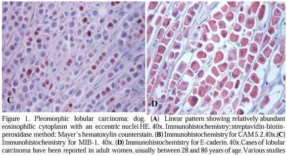
Tissue samples were collected for histopathologic examination from a 7-year-old female dog (mixed breed) with a nodule in the left abdominal and inguinal mammary glands that was brought to the surgery in the Porto Veterinary Hospital (Oporto-Portugal). The formalin-fixed tissue was embedded in paraffin, sectioned at 4mm and stained with haematoxylin and eosin (H&E) and PAS. For immunohistochemistry the streptavidin-biotin-peroxidase method was used in an automated system - Lab Vision (Cassali et al., 2001). Monoclonal antibodies for CAM 5.2 (Becton & Dickinson, USA) diluted 1:20, MIB-1 (Immunotech, France) diluted 1:30, progesterone receptor (Immunotech, France) diluted 1:40, E-caderin (Transduction Laboratories, USA) diluted 1:50 and also polyclonal antibodies for p53 (Novocastra, United Kingdom) and c-erbB2 ((Dako, Denmark)) were used.
Macroscopically the tumor mass was white, with a yellowish colour, and measured 15.0x7.0x7.0 cm and weighted 214.0 g. Microscopically, the tumor cells were loosely dispersed in the stroma or arranged in a linear pattern showing a relatively abundant eosinophilic cytoplasm and an eccentric nuclei (Fig. 1A). Many cells contained intracytoplasmic vacuolar dots which were PAS positive. Metastases were observed in the left and right inguinal lymph nodes. Immunohistochemical analyses showed positivity for CAM 5.2 (Fig. 1B), MIB-1 (44%) (Fig. 1C), and lack of expression for PR, p53 and c-erbB2. E-caderin expression was abnormal, with cytoplasmic staining without a membrane pattern (Fig. 1D).
The pleomorphic lobular carcinoma is defined as invasive with the infiltrating pattern of the classical lobular carcinoma, but with more pleomorphic nuclei, varying degrees of greater contour irregularity, increased mitotic activity and/or greater nuclear size (Eusebi et al., 1992; Rhadi, 2000). The recognition of this variant mammary tumor is very significant due to its a more aggressive behavior than the classical type an can be confused with invasive ductal carcinoma (Radhi, 2000).
Based on previous reports on invasive lobular carcinoma in the human species, the similar cytomorphologic and immunohistochemical features of dog mammary pleomorphic lobular carcinoma here described enabled the diagnosis of canine mammary gland primary pleomorphic lobular carcinoma. This is the first report of this type of mammary tumor in veterinary medicine.
ACKNOWLEDGEMENTS
This work was partially supported by grants from CAPES/ICCTI (035/98; 423 CAPES) and CNPq.
REFERENCES
Recebido para publicação em 16 de outubro de 2001
Recebido para publicação, após modificações, em 15 de maio de 2002
- CASSALI, G.D.; SILVA, P.; REMA, A. et al. A new methodology for the improvement of diagnostic immunohistochemistry in canine veterinary pathology: automated system using human monoclonal and polyclonal antibodies. Arq. Bras. Med. Vet. Zootec, v.53, p.326-331, 2001.
- Di COSTANZO, O.; ROSEN, P.P.; GAREEN, I. et al. Prognosis in infiltrating lobular carcinoma. An analysis of classical and variant tumors. Am. J. Surg. Pathol., v.14, p.12-23, 1990.
- EUSEBI, V.; MAGALHÃES, F.; AZZOPARDI, J.G. Pleomorphic lobular carcinoma of the breast: an aggressive tumor showing apocrine differentiation. Human Pathol., v.23, p.655-662, 1992.
- LEBEAU, A. L'age du chien et celui de l'homme. Assai de statistique sur la mortalité canine. Bul. Acad. Vet. France, v.26, p.229-232, 1953.
- RADHI, J.M. Immunohistochemical analysis of pleomorphic lobular carcinoma: higher expression of p53 and chromogranin and lower expression of ER and PgR. Histopathology, v.36, p.156-60, 2000.
- ROSEN, P.P. Rosen's breast pathology Philadelphia: Lippincott Raven, 1997. p. 545-562.
- ROSEN, P.P.; OBERMAN, H.A. Tumors of the mammary gland. Washington: Armed Forces Institute of Pathology, 1993. 390p.
Publication Dates
-
Publication in this collection
25 Mar 2003 -
Date of issue
Dec 2002
History
-
Reviewed
15 May 2002 -
Received
16 Oct 2001