Abstracts
The symptomatology and treatment of two dogs bitten by Crotalus durissus terrificus are described. Neurological signs were present few minutes after the accident with local anesthesia and ataxia of the affected limb and neurotoxic fascia. Alanine aminotransferase (ALT), aspartate aminotransferase (AST), creatinine kinase (CK), lactate dehydrogenase (LDH) and calcium were evaluated in an attempt to investigate muscle damage. Renal failure was not observed but some alterations were detected in urine. Urine density was low and the urinary sediment contained granular clumps and small round cells. Muscle samples were obtained from both legs for histopathological study, showing edema and isolated necrotic fibers. Both dogs received treatment within four hours after the accident by intravenous route. The antivenom was administered diluted in 250ml of Ringer solution in a dose enough to inactivate more than 8mg of venom. Dexamethasone was applied previously to the antivenom. Clinical evolution was good and both patients were in good health condition on the second day after the accident.
Dog; rattlesnake; Crotalus durissus terrificus; accident
Descreve-se a sintomatología e o tratamento de dois cães picados por cascavel da America do Sul (Crotalus durissus terrificus) na Argentina. Sinais neurológicos foram evidentes poucos minutos após o acidente, incluindo anestesia local, ataxia do membro afetado e fascia neurotóxica. As enzimas aminotransferase da alanina, aminotransferase do aspartato, quinase da creatinina, desidrogenase de lactato e o cálcio foram determinados para analisar dano muscular. Falha renal não foi observada, mas algunas alterações foram detectadas na urina, incluindo densidade baixa, sedimento com grumos granulares e células pequenas redondas. Biópsias de músculo foram obtidas de ambas as pernas para histopatología, revelando edema e fibras necróticas isoladas. Ambos os cães receberam tratamento quatro horas após o acidente, pela via intravenosa. A antitoxina foi administrada diluída em 250ml de solução Ringer lactato na dose capaz de neutralizar 8mg de toxina. Dexametasona foi aplicada antes de administrar a antitoxina. A evolução clínica foi boa e ambos os pacientes estavam com boa saúde no segundo dia após o acidente.
Cão; cascavel; Crotalus durissus terrificus; acidente
Case Report
[Relato de Caso]
American rattlesnake (Crotalus durissus terrificus) bite accidents in dogs in Argentina
[Acidente por mordida de cascavel (Crotalus durissus terrificus) em cães na Argentina]
P. Koscinczuk, O. Acosta de Pérez, P. Teibler, S. Maruñak, A.S. Rosciani
Facultad de Ciencias Veterinarias, UNNE
Sargento Cabral 2139
(3400) Corrientes, Argentina
Recebido para publicação, após modificações, em 28 de maio de 1999.
E-mail: patmed@vet.unne.edu.ar
ABSTRACT
The symptomatology and treatment of two dogs bitten by Crotalus durissus terrificus are described. Neurological signs were present few minutes after the accident with local anesthesia and ataxia of the affected limb and neurotoxic fascia. Alanine aminotransferase (ALT), aspartate aminotransferase (AST), creatinine kinase (CK), lactate dehydrogenase (LDH) and calcium were evaluated in an attempt to investigate muscle damage. Renal failure was not observed but some alterations were detected in urine. Urine density was low and the urinary sediment contained granular clumps and small round cells. Muscle samples were obtained from both legs for histopathological study, showing edema and isolated necrotic fibers. Both dogs received treatment within four hours after the accident by intravenous route. The antivenom was administered diluted in 250ml of Ringer solution in a dose enough to inactivate more than 8mg of venom. Dexamethasone was applied previously to the antivenom. Clinical evolution was good and both patients were in good health condition on the second day after the accident.
Keywords: Dog, rattlesnake, Crotalus durissus terrificus, accident
RESUMO
Descreve-se a sintomatología e o tratamento de dois cães picados por cascavel da America do Sul (Crotalus durissus terrificus) na Argentina. Sinais neurológicos foram evidentes poucos minutos após o acidente, incluindo anestesia local, ataxia do membro afetado e fascia neurotóxica. As enzimas aminotransferase da alanina, aminotransferase do aspartato, quinase da creatinina, desidrogenase de lactato e o cálcio foram determinados para analisar dano muscular. Falha renal não foi observada, mas algunas alterações foram detectadas na urina, incluindo densidade baixa, sedimento com grumos granulares e células pequenas redondas. Biópsias de músculo foram obtidas de ambas as pernas para histopatología, revelando edema e fibras necróticas isoladas. Ambos os cães receberam tratamento quatro horas após o acidente, pela via intravenosa. A antitoxina foi administrada diluída em 250ml de solução Ringer lactato na dose capaz de neutralizar 8mg de toxina. Dexametasona foi aplicada antes de administrar a antitoxina. A evolução clínica foi boa e ambos os pacientes estavam com boa saúde no segundo dia após o acidente.
Palavras-chave: Cão, cascavel, Crotalus durissus terrificus, acidente.
INTRODUCTION
Venomous animals have specialized glands to produce venom which acts immobilizing and killing their prey, beginning the digestive process (Hundelson & Hundelson, 1995). Envenoming caused by Crotalus durissus terrificus (South American rattlesnake) have low incidence in Argentina (Esteso, 1985) but the effects of floods during March and April, 1998, increased the number of accidents. Rattlesnake venom contains neurotoxic phospholipase A2. The toxic effects of this enzyme start a few hours after the patient is bitten, the most important occurring in cranial nerves III, IV and XI (Barraviera et al., 1993). Symptoms include palpebral ptosis, mydriasis, evident decreased muscular response, ophtalmoplegy and nystagmus. The only way to control these neurotoxic effects is the administration of specific antivenom within six hours of the envenomization (Esteso, 1985). These toxins also produce muscular necrosis (Gopalakrishnakone et al., 1984) and may cause injury to other organs such as the liver, as observed in a human patient with massive hepatic necrosis (Barraviera et al., 1989). Renal lesions with myoglobinuria are a consequence of the systemic myolisis and of the direct effect of the venom in the kidneys (Barraviera et al., 1993; Amorin et al., 1969).
CASE REPORT
Two naturally envenomized dogs by Crotalus durissus terrificus were referred to the Veterinary Hospital of the Faculty of Veterinary Medicine of Corrientes city, one a female and the other a male.
Blood samples were taken from cephalic vein after 5, 24, 48 and 168 hours of the accident. Colorimetric determinations of alanine aminotransferase (ALT), aspartate aminotransferase (AST) using the Reitman and Frankel method, and creatinine kinase (CK) which catalyzes the transference of a phosphate group of ATP to creatinine at pH 9 to produce creatininphosphate were performed. Free phosphorus as a result of the hydrolysis of creatininphosphate is proportional to the enzymatic activity and was determined by phosphatemia W colorimetric reaction from Wiener Lab. At the same time urea, creatinine and calcium serum values were studied.
Several urine samples were collected from each dog by cystocentesis or by free catch and processed within the first hour after to accident to avoid failures of collection. Chemical examination of urine included a Bayers assay for pH, protein, glucose, ketones, occult blood, and bilirubin. Specific gravity was evaluated by refractometry and the sediment was extended, fixed and coloured with hematoxiline-eosine for cytologic analysis.
Biopsies for histopathology were performed in only one animal. Muscle samples were obtained from both posterior limbs, the affected and the contralateral one. Then they were fixed with Bouin and processed by classical histologic technique.
The symptomatology was similar in both patients. The skin area where the venom was injected could not be visually identified. Neurological signs were present a few minutes after the accident as a local hypoalgesia. After the hypoalgesia, the affected limb suffered anesthesia and ataxia. After one hour the opposite leg showed a similar symptomatology. Neurotoxic fascia was present 30 minutes after the accident with palpebral ptosis, anisocoria, sialorrhea and with altered esophagosalivary reflex (Fig. 1). Previous to the fluid therapy, an urine test was performed, revealing pH 7, with a density of 1.010 and proteinuria. Hemoglobinuria was detected perhaps because of the myoglobinuria.
Bites produced by venomous snakes require critical assessment for systemic signs. In both dogs fluid therapy with lactated Ringers solution was initiated after the diagnosis had been established. In an attempt to avoid the shock a corticosteroid was administered (dexamethasone 4,4mg/kg) (Tilley et al., 1998). After the corticosteroid a vial of the specific antivenom was administered to neutralize 8mg of venom.
After the second day, special care was taken with food which was given as small pellets in deep dishes, because of the loss of esophagosalivary reflex.
After initiating the treatment, respiratory distress disappeared and two hours later also the palpebral ptosis. Hiperalgia appeared on the second day, and the esophagosalivary reflex was altered during three days.
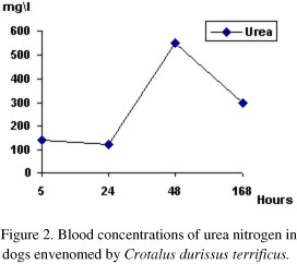
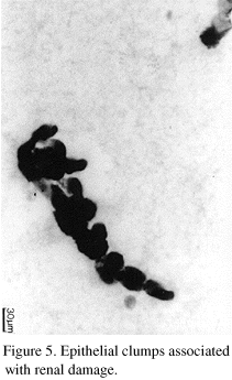
Blood chemistry revealed an increase in the urea nitrogen level which was the highest within 48 hours (Fig. 2), but creatinine did not show any variation. AST and CPK increased after the first four hours, reaching the highest value at 24 hours (Fig. 3). Urine chemical tests showed pH of 7.0, density of 1.010 and presence of protein and hemoglobin. This hemoglobin was not differenciated from mioglobin, but it could be suspected because of the muscle damage. In the sediment there were round cells with picnotic nucleous, probably of tubular origin, among vaginal epithelial cells (Fig. 4). Necrotic epithelial clumps could be associated with tubular damage (Fig. 5). In spite of the renal lesions, which were suggested by the urine test, there was not renal failure, and the values of urea nitrogen and creantinine were normal after the first week.
The biopsies performed from the injected muscle showed markedly edema of conective tissue and there were many muscle fibers which underwent necrosis. In the contralateral limb occasional myonecrosis could be observed interspersed among normal muscle fibers (Fig. 6).
DISCUSSION
The venom of Crotalus durissus terrificus cause flaccid paralysis in laboratory animals, and death by respiratory failure (Brazil, 1980). It happens because crotoxin inhibit the neuromuscular transmission in vertebrates (Hawgood & Smith, 1977). In this work, it was observed both dogs with hyporreflexia and incoordination motion leading them to be lying.
The venom neurotoxicity was evident a few minutes after the accident. The symptomatology was similar to the alteration of cranial nerves III, IV and XI (Barraviera et al., 1993). These alterations produce the neurotoxic fascie of a rattlesnake accident.
When biopsy was performed, muscular necrosis was observed, not only where the injection was performed but in the contralateral muscle as well. Any snake envenoming that causes muscular necrosis could cause the release of myoglobin (Magalhães et al., 1980). Differentiating myoglobin from hemoglobin in the urine may be difficult without readily available methods. Patients had got hemoglobin positive in the urine test, but it was suspected a problem of myonecrosis. The kidneys alterations observed in man (Azevedo Marques et al., 1985; 1987) with tubular obstruction with clumps are similar to those found in this work.
The presence of altered muscle fibers in the contralateral limb, showed the systemic action of some venom components. This could be supported by the urea and the AST, CPK values, all involved with the muscle metabolism.
- AMORIN, M.F., MELLO, R.F., SALIBA, F. Lesőes renais induzidas experimentalmente no căo pelo veneno crotálico. Mem. Inst. Butantan, v.24, p.281, 1969.
- AZEVEDO-MARQUES, M.M., CUPO, P., COIMBRA, T.M. et al. Myonecrosis, myoglobinuria and acute renal failure induced by South American rattlesnake (Crotalus durissus terrificus) envenomation in Brazil. Toxicon, v.23, p.631-636,1985.
- AZEVEDO-MARQUES, M.M., HERING, S.E., CUPO, P. Evidence that Crotalus durissus terrificus (South American rattlesnake) envenomation in human causes myolisis rather than hemolysis. Toxicon, v.11, p.1162-1168, 1987.
- BARRAVIERA, B., BONJORNO Jr. J.C., ARAKAKI, D. et al. A retrospective study of 40 victims of Crotalus snake bites. Analysis of the hepatic necrosis observed in one patient. Rev. Soc. Bras. Med. Trop, v.22, p.5-12,1989.
- BARRAVIERA, B., AGAPEJEV, S., VEIGA CASTRO, C. Acidente crotálico: importância do diagnóstico clinico. ABP Supl. Bras. Med., v.67, p.36-37,1993.
- BRAZIL, O.V. Venenos ofídicos neurotóxicos. Rev. Ass. Med. Bras, v.26, p.212-218, 1980.
- ESTESO, S.C. Ofidismo en la República Argentina Argentina: ARPON, 1985.170p.
- GOPALAKRISHNAKONE, P., DEMPSTER, D.W., HAWGOOD, B.J. et al. Cellular and mitochondrial changes induced in the structure of murine skeletal muscle by crotoxin, a neurotoxic phospholipase A2 complex. Toxicon, v.22, p.85, 1984.
- HAWGOOD, B.J., SMITH, J.W. The mode of action at the mouse neuromuscular junction of the phospholipase A crotopotin complex isolated from the venom of the South American rattlesnake. Br. J. Pharmacol, v.61, p.597-606, 1977.
- HUNDELSON, S., HUNDELSON, P. Pathophysiology of snake envenomation and evaluation of treatment. Part II. Comp. Small Anim, v.17, p.889-897, 1995.
- MAGALHĂES, R.A., RIBEIRO, M.F., REZENDE, N.A. et al. Rabdomiólisis secundária a acidente ofidico crotálico (Crotalus durissus terrificus). Rev. Inst. Med. Trop., v.28, p.315-321, 1980.
- TILLEY, L.T., SMITH, F.W.K., MAC MURRAY, A.C. La consulta veterinaria en 5 minutos. Argentina: Intermédica, p.1201, 1998.
Publication Dates
-
Publication in this collection
14 Aug 2000 -
Date of issue
Apr 2000
History
-
Received
28 May 1999