Abstract
Descrevem-se os achados citomorfológicos de um tumor maligno de mama em uma cadela Poodle de sete anos de idade, o qual foi observado inicialmente pelo exame citológico do derrame pleural. Comparam-se os aspectos citológicos do derrame pleural e punção aspirativa com agulha fina do tumor com aqueles descritos para o câncer de mama na espécie humana.
Cão; citologia; derrame pleural; tumor de mama
Dog; cytology; pleural effusion; mammary tumor
Cão; citologia; derrame pleural; tumor de mama
COMMUNICATION
(Comunicação)
Cytological diagnosis of a metastatic canine mammary tumor in pleural effusion(Diagnóstico citológico de tumor mamário metastático canino em derrame pleural)
G.D. Cassali1, F. Gärtner2, M.J. Vieira da Silva3, F.C. Schmitt2
1Caixa Postal 486
31270-901 Belo Horizonte, MG - Brasil
2Instituto de Patologia e Imunologia Molecular da
Universidade do Porto (IPATIMUP), Porto Portugal
3Clínica Veterinária Matosinhos, Matosinhos Portugal Recebido para publicação em 27 de novembro de 1998.
e-mail: cassalig@mono.icb.ufmg.br
The involvement of the serous cavities by malignant neoplasms has important therapeutic and prognostic implications. In most instances, malignant cells are found during the course of the disease in effusions from patients with a known history of malignant neoplasm. In some patients, however, a malignant effusion is the first indication of cancer (Monte et al., 1987).
The breast is the most common source of malignant pleural effusions and the second most common source of malignant ascites in women. In most instances, the diagnosis of mammary cancer is established before pleural metastases occur; rarely does an occult carcinoma present initially with a malignant effusion (Monte et al., 1987).
In human species the cytologic examination of effusions to determine the presence of malignant cells has been done since 1867 (Lucke & Klebs, 1867 apud Sears & Hadju, 1987). However we are not aware of description of malignant cells in effusions in the veterinary literature. Herein cytomorphologic features of a malignant canine mammary tumor which was observed initially by the cytologic examination of a pleural effusion, is described.
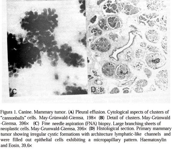
A Poodle bitch aged seven years presented with a history of cardiac failure with pleural effusion for approximately 1 year. Ascitic effusions have also developed in the last 3 months. Pleural effusions have been collected; the slides were made by direct smearing sediment of the centrifuged specimen. The material obtained was smeared on to a glass slide and either air dried for May-Grünwald-Giemsa staining or 95% ethanol fixed for Papanicolaou staining. Microscopic examination exhibited an abundant number of isolated neoplastic epithelial cells and clusters of "cannonballs" cells, with an increased nucleus/cytoplasm ratio, compatible with the diagnosis of adenocarcinoma (Fig. 1A, B)
A putative mammary origin of a primary tumor was considered and verified by the clinical examination of cranial and caudal thoracic left glands. A fine needle aspiration (FNA) biopsy was performed using 0.6mm of diameter needle attached to a 10ml syringe held by a standard metal syringe holder. The material obtained was smeared on to a glass slide and either air dried for May-Grünwald-Giemsa staining or 95% ethanol fixed for Papanicolaou staining.
The aspirates were highly cellular with numerous large branching sheets of neoplastic cells as well as isolated round to oval cells. The nuclei had irregular distributed chromatin with single or multiple nucleoli (Fig. 1C). The cytological findings were compatible with the diagnosis of adenocarcinoma.
Thirty days after the diagnosis of a mammary cancer, the owner decided to sacrifice the animal. Ascitic effusions have been collected and the same cytological aspects verified in pleural effusions were found. A tumor mass weighting 52 grams and measuring 11.5 ´ 5.0 ´ 1.5 cm was found in the cranial and caudal thoracic left glands. No other tumor mass was detected in other organs, but in the right kidney was found an infarction measuring 1.2 ´ 0.7cm. Formalin fixed tissue tumor mass and specimens of the other organs were paraffin embedded and sections 4mm thick were cut and stained with Haematoxylin and Eosin (HE).
Histologically the mammary tumor was characterized by the presence of numerous irregular cystic formations, distributed diffusely through the mammary parenchyma tissue. Some of these cystic spaces had lymphatic-like channels and were filled out with epithelial cells exhibiting a micropapillary pattern (Fig. 1D). The epithelial cells showed large eosinophilic cytoplasm and vesicular pleomorphic nucleus with prominent nucleoli. Numerous mitotic figures were depicted. The histological findings were compatible with the diagnosis of invasive micropapillary mammary carcinoma. Micrometastasis were observed in lymph nodes, lung, epicardium, pericardium and kidneys. In the right kidney neoplastic emboli were verified in the cortical-medullar arteries with coagulative necrosis.
Cytologic results of the pleural effusions in this case were similar to the ones described in the literature. In human species breast carcinomas in pleural effusions are often present in cell clusters, with more than 80 percent of the sample forming groups of relatively uniform cells in three-dimensional arrangements that have smooth outer-contours. Multinucleation, macronucleoli, cytoplasmic vacuolization or the presence of bizarre cells occur less frequently than in ovarian or lung cancer (Sheibani & Esteban, 1997; Silverman, 1997).
The cytological features of this case obtained by fine needle aspiration are the same observed in human species and were compatible with malignancy, i.e. cellular smears, loosely cohesive and individual scattered malignant cells and malignant epithelial cells arranged in three-dimensional clusters (Naylor, 1997).
In humans, cytologic examination of a serous fluid is of paramount importance because the finding of cancer cells in such a specimen denotes that the patient has cancer that is not only advanced but almost always incurable (Naylor, 1997). These results showed a resemblance between human and dog mammary cancer and demonstrate the relevance of pleural effusion examination in canine with a mammary gland tumor.
ACKNOWLEDGMENTS
We are grateful to Raquel Soares for critically reviewing the English manuscript and FAPEMIG (Grant n° CBS838/96) and CAPES/ICCTI (Grant n° 035/98) for finnancial support.
RESUMO
Descrevem-se os achados citomorfológicos de um tumor maligno de mama em uma cadela Poodle de sete anos de idade, o qual foi observado inicialmente pelo exame citológico do derrame pleural. Comparam-se os aspectos citológicos do derrame pleural e punção aspirativa com agulha fina do tumor com aqueles descritos para o câncer de mama na espécie humana.
Palavras-Chave: Cão, citologia, derrame pleural, tumor de mama
- MONTE, S.A., EHYA, H., LANG, W. R. Positive effusion cytology as the initial presentation of malignancy. Acta Cytol, v.31, p.448-452, 1987.
- NAYLOR, B. Pleural, peritoneal, and pericardial fluids. In: BIBBO, M. (Ed.) Comprehensive cytopathology, 2.ed., Philadelphia: W.B Saunders, 1997. p.551-621.
- SEARS, D., HADJU, S.I. The cytologic diagnosis of malignant neoplasms in pleural and peritoneal effusions. Acta Cytol, v.31, p.85-97, 1987.
- SHEIBANI, K, ESTEBAN, J. Pleura, pericardium, and peritoneum. In: SILVERBERG, S. G., DeLELLIS, R.A., FRABLE, W.J. (Eds.) Principles and practice of surgical pathology and cytopathology 3.ed. New York: Churchill Livingstone, 1997. v.2, p.1299-1348.
- SILVERMAN, J.F. Breast. In: BIBBO, M. (Ed.) Comprehensive cytopathology, 2.ed., Philadelphia: W.B Saunders, 1997. p.731-780.
Publication Dates
-
Publication in this collection
17 Apr 2001 -
Date of issue
Aug 1999
History
-
Received
27 Nov 1998