Abstract
Semiconductor laser devices are readily available and practical radiation sources providing wavelength tenability and high monochromaticity. Low-intensity red and near-infrared lasers are considered safe for use in clinical applications. However, adverse effects can occur via free radical generation, and the biological effects of these lasers from unusually high fluences or high doses have not yet been evaluated. Here, we evaluated the survival, filamentation induction and morphology of Escherichia coli cells deficient in repair of oxidative DNA lesions when exposed to low-intensity red and infrared lasers at unusually high fluences. Cultures of wild-type (AB1157), endonuclease III-deficient (JW1625-1), and endonuclease IV-deficient (JW2146-1) E. coli, in exponential and stationary growth phases, were exposed to red and infrared lasers (0, 250, 500, and 1000 J/cm2) to evaluate their survival rates, filamentation phenotype induction and cell morphologies. The results showed that low-intensity red and infrared lasers at high fluences are lethal, induce a filamentation phenotype, and alter the morphology of the E. coli cells. Low-intensity red and infrared lasers have potential to induce adverse effects on cells, whether used at unusually high fluences, or at high doses. Hence, there is a need to reinforce the importance of accurate dosimetry in therapeutic protocols.
DNA; Escherichia coli; Filamentation; Laser
Introduction
Low-intensity lasers are lightweight, available sources of monochromatic non-ionizing radiation (11. Niemz MH. Laser-tissue interactions: Fundamentals and applications. New York: Springer-Verlag; 2007.). Because they are practical and low-cost, these devices are increasingly being used in health care. In nonphotosynthetisizing cells, laser light absorption occurs via chromophores and alterations in cell physiology have been reported (22. Karu T. Primary and secondary mechanisms of action of visible to near-IR radiation on cells. J Photochem Photobiol B 1999; 49: 1–17, doi: 10.1016/S1011-1344(98)00219-X.
https://doi.org/10.1016/S1011-1344(98)00...
). Chromophores, which act as intracellular photoacceptors, are responsible for the biological effects of low-intensity lasers (33. Karu TI, Lyapunova TS, Pomoshnikova NA. The activation of yeast metabolism with He-Ne laser radiation. IV. Relationship between the activity of catalase and protein synthesis. Lasers Life Sci1993; 5: 251–258.). Certain reaction centers in cytochrome c oxidase (Cua and Cub or hemes a and a3) in mammalian cells and cytochrome bd and bo complexes in Escherichia coli cells have been described as the main cellular photoacceptors (33. Karu TI, Lyapunova TS, Pomoshnikova NA. The activation of yeast metabolism with He-Ne laser radiation. IV. Relationship between the activity of catalase and protein synthesis. Lasers Life Sci1993; 5: 251–258.). After absorption of laser radiation energy at low fluences by such photoacceptors, transduction processes are responsible for activating intracellular signaling pathways, thereby amplifying the primary photosignal (44. Karu T, Pyatibrat L. Gene expression under laser and light-emitting diodes radiation for modulation of cell adhesion: Possible applications for biotechnology. IUBMB Life 2011; 63: 747–753.). Highly reactive chemical species (i.e., reactive oxygen and nitrogen species) are involved in the transduction processes where they function as second messages, interact with biomolecules, and alter cellular functions and gene expression (44. Karu T, Pyatibrat L. Gene expression under laser and light-emitting diodes radiation for modulation of cell adhesion: Possible applications for biotechnology. IUBMB Life 2011; 63: 747–753.,55. Hawkins DH, Abrahamse H. The role of laser fluence in cell viability, proliferation, and membrane integrity of wounded human skin fibroblasts following helium-neon laser irradiation. Lasers Surg Med 2006; 38: 74–83, doi: 10.1002/lsm.20271.
https://doi.org/10.1002/lsm.20271...
). It is possible that photobiological side-effects occur when the antioxidant systems are not capable of protecting the cells against free radical attack. This situation can occur when antioxidant systems are not functioning, or when inadequate exposure to low-intensity lasers at high doses arises. An intracellular imbalance between oxidant and antioxidant contents means that free radicals might occur in cells exposed to low-intensity lasers when high doses are used. At therapeutic doses, sub-lethal DNA damage has been reported after exposure to low-intensity red and infrared lasers in eukaryotic (55. Hawkins DH, Abrahamse H. The role of laser fluence in cell viability, proliferation, and membrane integrity of wounded human skin fibroblasts following helium-neon laser irradiation. Lasers Surg Med 2006; 38: 74–83, doi: 10.1002/lsm.20271.
https://doi.org/10.1002/lsm.20271...
66. Godon C, Cordelieres FP, Biard D, Giocanti N, Megnin-Chanet F, Hall J, et al. PARP inhibition versus PARP-1 silencing: different outcomes in terms of single-strand break repair and radiation susceptibility. Nucleic Acids Res2008; 36: 4454–4464, doi: 10.1093/nar/gkn403.
https://doi.org/10.1093/nar/gkn403...
77. Mbene AB, Houreld NN, Abrahamse H. DNA damage after phototherapy in wounded fibroblast cells irradiated with 16 J/cm(2). J Photochem Photobiol B 2009; 94: 131–137, doi: 10.1016/j.jphotobiol.2008.11.002.
https://doi.org/10.1016/j.jphotobiol.200...
) and prokaryotic cells (89. Kohli R, Gupta PK. Irradiance dependence of the He-Ne laser-induced protection against UVC radiation in E. coli strains. J Photochem Photobiol B 2003; 69: 161–167.,99. Fonseca AS, Geller M, Bernardo FM, Valenca SS, de PF. Low-level infrared laser effect on plasmid DNA. Lasers Med Sci 2012; 27: 121–130, doi: 10.1007/s10103-011-0905-2.
https://doi.org/10.1007/s10103-011-0905-...
).
Although low-intensity laser radiation can potentially damage DNA, therapeutic protocols based on it are used successfully to improve wound healing (1010. Peplow PV, Chung TY, Baxter GD. Laser photobiomodulation of wound healing: a review of experimental studies in mouse and rat animal models. Photomed Laser Surg 2010; 28: 291–325, doi: 10.1089/pho.2008.2446.
https://doi.org/10.1089/pho.2008.2446...
), accelerate the repair of skin, cartilage and bone, to treat nerve injuries and relieve inflammation (1111. Maia ML, Bonjardim LR, Quintans JS, Ribeiro MA, Maia LG, Conti PC. Effect of low-level laser therapy on pain levels in patients with temporomandibular disorders: a systematic review. J Appl Oral Sci 2012; 20: 594–602, doi: 10.1590/S1678-77572012000600002.
https://doi.org/10.1590/S1678-7757201200...
) and pain (1212. Gross AR, Dziengo S, Boers O, Goldsmith CH, Graham N, Lilge L, et al. Low level laser therapy (LLLT) for neck pain: a systematic review and meta-regression. Open Orthop J 2013; 7: 396–419, doi: 10.2174/1874325001307010396.
https://doi.org/10.2174/1874325001307010...
). The scientific basis of laser applications in therapy is the so-called biostimulation (or biomodulation) effect, which results from alterations of intracellular processes, mainly via an increase in metabolism and the rate of cell division (22. Karu T. Primary and secondary mechanisms of action of visible to near-IR radiation on cells. J Photochem Photobiol B 1999; 49: 1–17, doi: 10.1016/S1011-1344(98)00219-X.
https://doi.org/10.1016/S1011-1344(98)00...
).
The biological effects of low-intensity lasers are dependent on the exposure parameters used. Energy densities, directionality, high monochromaticity and emission mode properties are characteristics that enable semiconductor laser devices to treat various diseases, and the different clinical protocols suggested for their use can be found in specialized literature on this topic (1111. Maia ML, Bonjardim LR, Quintans JS, Ribeiro MA, Maia LG, Conti PC. Effect of low-level laser therapy on pain levels in patients with temporomandibular disorders: a systematic review. J Appl Oral Sci 2012; 20: 594–602, doi: 10.1590/S1678-77572012000600002.
https://doi.org/10.1590/S1678-7757201200...
) and in guides on laser devices. These protocols are based on low-energy densities (fluences) or low-power densities and for this reason low-intensity lasers are considered safe for clinical applications. Also, red and near-infrared radiation (600 up to 1300 nm) is not considered to induce significant adverse effects in biological tissues (22. Karu T. Primary and secondary mechanisms of action of visible to near-IR radiation on cells. J Photochem Photobiol B 1999; 49: 1–17, doi: 10.1016/S1011-1344(98)00219-X.
https://doi.org/10.1016/S1011-1344(98)00...
), unlike ultraviolet radiation, which induces hyperpigmentation, aging and carcinogenesis (1313. Amaro-Ortiz A, Yan B, D'Orazio JA. Ultraviolet radiation, aging and the skin: prevention of damage by topical cAMP manipulation. Molecules 2014; 19: 6202–6219, doi: 10.3390/molecules19056202.
https://doi.org/10.3390/molecules1905620...
). Under low fluences (0.1 up to 100 J/cm2), low-intensity lasers are considered to generate nonthermal and nondestructive effects (11. Niemz MH. Laser-tissue interactions: Fundamentals and applications. New York: Springer-Verlag; 2007.). However, high energy densities and intensities are deposited in a small volume and over a short time period, thereby delivering high-dose radiation to the biological tissue exposed to such lasers. Hence, the clinical outcomes of laser use depend on delivery of accurate doses of laser radiation and ensuring that adverse effects cannot occur through accidental high-dose exposure. However, few experimental studies on the biological effects induced by low-intensity lasers at unusual doses exist, making research in this area important as undesirable effects from low-dose lasers can occur via accidental exposure or when non-calibrated devices are used. Therefore, the work presented here investigated the survival, filamentation induction and morphology of E. coli cells deficient in repair of oxidative DNA lesions when exposed to low-intensity red and infrared laser radiation at unusually high fluences.
Material and Methods
Low-intensity red and near-infrared lasers
Therapeutic low-intensity red and near-infrared lasers (Photon Lase III) were purchased from DMC Equipamentos Ltda. (Brazil). The laser parameters are shown in Table 1.
E. coli cell survival
Cultures of E. coli AB1157 (wild-type), JW1625-1 (deficient in endonuclease III) and JW2146-1 (deficient in endonuclease IV) were exposed to low-intensity red and infrared lasers and their survival rates were evaluated. From stocks in stationary growth phase, cultures of these strains were prepared to attain their exponential growth phase (i.e., 108 cells/mL; 2–3 h, 37°C). Other experiments were carried out with cultures of the same E. coli strains in the stationary growth phase (1010 cells/mL; 18 h, 37°C). Bacterial cells were centrifuged twice (700 g, 15 min) and resuspended in saline (0.9% NaCl) each time. Aliquots (50 µL, n=5, for each fluence) of the bacterial suspensions (108 cells/mL) were exposed, at room temperature and under white light (fluorescent lamps), to low-intensity red and infrared lasers. The exposure time of the cells was automatically adjusted by the laser device as a function of the fluence. The laser device was positioned such that almost all the surface of the bacterial aliquot suspension was covered by the laser beam. Controls were bacterial suspensions not exposed to lasers. Immediately after exposure to a laser, the bacterial suspensions were diluted in normal saline and spread onto Petri dishes containing solidified rich medium (1.5% agar). Bacterial colonies were counted after incubation (37°C, 18 h) and the survival fractions were calculated (1414. Fonseca AS, Moreira TO, Paixao DL, Farias FM, Guimaraes OR, de PS, et al. Effect of laser therapy on DNA damage. Lasers Surg Med 2010; 42: 481–488, doi: 10.1002/lsm.v42:6.
https://doi.org/10.1002/lsm.v42:6...
).
Bacterial filamentation assays
To evaluate filamentation induction, exponential and stationary E. coli AB1157, JW1625-1, and JW2146-1 cultures were obtained and exposed to low-intensity red and infrared lasers as described in the bacterial survival assay. Bacterial suspensions not exposed to lasers were used as controls. Immediately after exposure, aliquots (20 µL) were withdrawn, spread onto microscopic slides and stained by the Gram method (1515. Cappuccino JG, Sherman N. Microbiology: a laboratory manual. California: Benjamin Cummings Science Publishing; 1999.). Bacterial cells were visualized using a Carl Zeiss Axio Scope A1 microscope (Germany) equipped with an A-plan 40/0.65 objective, a 0.90 condenser and a 100W halogen lamp. The images were captured with an AxioCam HRc Sony 12M color microscopy camera (Carl Zeiss), using AxioVision software. Thereafter, the images were analyzed by Image-Pro Plus 6.0 software for Windows XP (Media Cybernetics, Inc., USA) to determine the bacterial filamentation percentages. A bacterial filament was considered to be 2.5 times the average area of a bacterial cell. Experiments were carried out in duplicate and the results represent the mean of three independent assays.
Bacterial morphological measurements
Bacterial suspensions of E. coli AB1157, JW2146-1, and JW1625-1 (108 cells/mL) were exposed to low-intensity red and infrared lasers as described above in the bacterial survival and filamentation assay methods. Immediately after laser exposure, aliquots were spread onto microscopic slides and stained by the Gram method (1515. Cappuccino JG, Sherman N. Microbiology: a laboratory manual. California: Benjamin Cummings Science Publishing; 1999.). Bacterial cells were visualized by light microscopy (300 cells for each laser exposure), as described in the bacterial filamentation assay method.
Statistical analysis
Data are reported as means±SD of the bacterial survival fractions, the bacterial filament percentages, and the surface area of the bacterial cells. One-way analysis of variance (ANOVA) was performed to verify potential statistical differences, followed by the Tukey post-test with P<0.05 indicating statistical significance. InStat software for Windows XP (GraphPad Software, USA) was used to perform the statistical analyses.
Results
Survival of E. coli cultures exposed to low-intensity red and infrared lasers
The survival fractions of exponentially grown E. coli AB1157, JW1625-1 and JW2146-1 cultures exposed to low-intensity red and infrared lasers are reported in Table 2. The data in this table show that exposure to these lasers did not significantly alter the survival fractions of the E. coli AB1157 and JW1625-1 cultures. However, red and infrared lasers significantly (P<0.05) decreased the survival fractions of JW2146-1 at the higher fluence (1000 J/cm2) evaluated herein.
The survival rates of stationary cultures of the same E. colistrains were evaluated to verify whether the low-intensity red and infrared laser effects are dependent on the physiological conditions of the cells (Table 3). Stationary E. coli AB1157 cultures had survival fractions similar to those of the exponential cultures. However, E. coli JW1625-1 had an increased survival fraction after exposure to red laser at the higher fluence level. No significant alteration of the survival fraction was obtained for E. coli JW1625-1 after infrared laser exposure. In contrast to the decreased survival fractions of the exponential cultures of E. coli JW2146-1, the survival fractions of stationary JW2146-1 cultures were not significantly modified by exposure to low-intensity red and infrared lasers.
Filamentation induction in E. coli cultures exposed to low-intensity red and infrared lasers
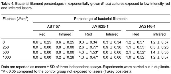
Figure 1 shows a photograph of representative cells from E. coli AB1157 cultures during the exponential growth phase (1A). Figure 1Bshows the bacterial cell image analysis. The bacterial filament percentages in exponential phase E. coli cultures are shown in Table 4. Data in this table show that the red and infrared lasers did not significantly induce the filamentation phenotype in E. coli AB1157. Also, infrared laser treatment did not significantly induce filament formation in E. coli JW1625-1 and JW2146-1 cultures. However, in the JW1625-1 cultures, exposure to low-intensity red laser significantly (P<0.05) induced an increase in the percentage of bacterial filaments, but in JW2146-1 cultures this effect was significant only at mid fluence (500 J/cm2).
Representative images of bacterial filamentation from AB1157 cultures in the stationary growth phase. A, Arrow denotes bacterial filamentation; B, same image illustrating how the image analysis was performed. A bacterial filament was considered to be present in a bacterium when the area of the bacterial cell was 2.5-times larger than the mean value of the area.
Stationary E. coli cultures were also exposed to red and infrared lasers to evaluate filamentation induction (Table 5). Similar to the results observed with the exponential cultures, E. coli AB1157 exposure to red laser treatment did not induce significant filamentation, but exposure to infrared laser at the higher fluence (1000 J/cm2) increased the level of this phenotype. In contrast to the results of the exponential cultures, red laser exposure did not induce significant filamentation in stationary E. coli JW1625-1. Interestingly, laser exposure significantly (P<0.05) reduced the filament percentage in stationary E. coli JW2146-1, except at 500 J/cm2 (no significant alteration) and at 1000 J/cm2 where a significant (P<0.05) increase in bacterial filaments was seen.
Effect of low-intensity red and infrared lasers on the surface area of E. coli cells
The surface area of individual E. coli cells was evaluated after exposure to lasers at high fluences (Tables 6 and 7). The data in Table 6 show that exposure to red and infrared lasers significantly (P<0.05) increased the surface area of exponential E. coli AB1157 cells. However, exposure to the red laser did not induce significant alteration of the surface area of E. coli JW1625-1 cells; infrared laser exposure at the highest fluences (500 and 1000 J/cm2), however, did reduce the surface area of these cells. No significant alterations to the surface area of E. coli JW2146-1 exposed to red and infrared lasers were observed.
To verify whether stationary cells, which differ physiologically to exponential cells, could be altered by laser exposure, the surface area of these cells was also evaluated. The data in Table 7 show that low-intensity red and infrared lasers did not induce significant alterations to the surface areas of stationary E. coli AB1157, JW1625-1, and JW2146-1 cells.
Discussion
Some clinical protocols based on low-intensity red and infrared lasers were proposed by empirical professional practice. Also, current device guides do not contain recommendations that prevent medical professionals from delivering high radiation doses to patients during laser exposure. Despite some discrepancies, experimental studies have mostly described the positive effects induced by such lasers when used at therapeutic doses. However, the effects of accidental exposure to high-dose radiation or exposure to non-calibrated laser devices have been neglected. Our research shows that, at high fluences, low-intensity red and infrared laser radiation used to treat soft tissue diseases can induce lethal effects on E. coli cultures, depending on the DNA repair mechanisms of the strain and the physiological condition of the cells (i.e., stationary or exponentially growing cells) (Tables 2 and 3). In previous studies, we have shown that E. coli (AB1157) cells proficient in DNA repair mechanisms are resistant to red and infrared laser exposure at fluences or doses in the therapeutic range (1616. Sergio LPS, Marciano RS, Polignano GAC, Guimarães OR, Geller M, Paoli F, et al. Evaluation of DNA damage induced by therapeutic low-level red laser. Clin Exp Dermatol Res 2012; 3: 166, doi: 10.4172/2155-9554.1000166.
https://doi.org/10.4172/2155-9554.100016...
1717. Canuto KS, Sergio LPS, Marciano RS, Guimarães OR, Polignano GAC, Geller M, et al. DNA repair in bacterial cultures and plasmid DNA exposed to infrared laser for treatment of pain. Laser Phys Lett 2013; 10: 065606, doi: 10.1088/1612-2011/10/6/065606.
https://doi.org/10.1088/1612-2011/10/6/0...
1818. Teixeira GR, Marciano RS, Sergio LPS, Polignano GAC, Guimarães OR, Geller M, et al. Effects of infrared laser at fluences used for treatment of dentin hypersensitivity on DNA repair in Escherichia coli and plasmids. Opt Laser Technol 2014; 64: 46–52, doi: 10.1016/j.optlastec.2014.04.023.
https://doi.org/10.1016/j.optlastec.2014...
). The data in Tables 2 and 3 suggest that these cells are also not inactivated by red and infrared laser exposure at high fluences. However, high fluences of red and infrared lasers decrease the survival rates of exponential phase E. coli cultures deficient in endonuclease III (JW1625-1) and E. coli cultures deficient in endonuclease IV (JW2146-1) (Table 2). As part of its base excision repair mechanism, endonuclease III repairs apurinic/apyrimidinic sites and damaged pyrimidines (1919. Romano CA, Sontz PA, Barton JK. Mutants of the base excision repair glycosylase, endonuclease III: DNA charge transport as a first step in lesion detection. Biochemistry 2011; 50: 6133–6145, doi: 10.1021/bi2003179.
https://doi.org/10.1021/bi2003179...
,2020. Kato S, Hashiguchi K, Igarashi K, Moriwaki T, Yonekura S, Zhang-Akiyama QM. Structural and functional properties of CiNTH, an endonuclease III homologue of the ascidian Ciona intestinalis: critical role of N-terminal region. Genes Genet Syst 2012; 87: 115–124, doi: 10.1266/ggs.87.115.
https://doi.org/10.1266/ggs.87.115...
). Similarly, endonuclease IV acts on apurinic/apyrimidinic sites and oxidatively damaged bases in E. coli (2121. Ramotar D. The apurinic-apyrimidinic endonuclease IV family of DNA repair enzymes. Biochem Cell Biol 1997; 75: 327–336, doi: 10.1139/o97-046.
https://doi.org/10.1139/o97-046...
2222. Kerins SM, Collins R, McCarthy TV. Characterization of an endonuclease IV 3′-5′ exonuclease activity. J Biol Chem 2003; 278: 3048–3054, doi: 10.1074/jbc.M210750200.
https://doi.org/10.1074/jbc.M210750200...
2323. Christov PP, Banerjee S, Stone MP, Rizzo CJ. Selective Incisincision of the α-N5-methyl-formamidopyrimidine anomer by Escherichia coli endonuclease. J Nucleic Acids2010; 2010: 850234.
2424. Mol CD, Hosfield DJ, Tainer JA. Abasic site recognition by two apurinic/apyrimidinic endonuclease families in DNA base excision repair: the 3′ ends justify the means. Mutat Res 2000; 460: 211–229, doi: 10.1016/S0921-8777(00)00028-8.
https://doi.org/10.1016/S0921-8777(00)00...
). Data obtained in our study indicated that sub-lethal oxidative lesions in DNA are induced in cells exposed to low-intensity red and infrared lasers, and that the survival of cells with failing DNA repair mechanisms decreased when exposed to such radiation. However, stationary endonuclease IV-deficient cells are not sensitive to lasers and the viability of endonuclease III-deficient cells is increased by red laser exposure at the higher fluence level we evaluated (1000 J/cm2). These results suggest that both endonuclease III- and endonuclease IV-deficient E. coli cells respond to low-intensity lasers depending on their physiological condition. Nevertheless, laser-induced effects on endonuclease III-deficient cells at high fluence might be related to an increase or acceleration of cellular proliferation (biostimulation or biomodulation effect) (11. Niemz MH. Laser-tissue interactions: Fundamentals and applications. New York: Springer-Verlag; 2007.,1717. Canuto KS, Sergio LPS, Marciano RS, Guimarães OR, Polignano GAC, Geller M, et al. DNA repair in bacterial cultures and plasmid DNA exposed to infrared laser for treatment of pain. Laser Phys Lett 2013; 10: 065606, doi: 10.1088/1612-2011/10/6/065606.
https://doi.org/10.1088/1612-2011/10/6/0...
) despite this effect not being observed at the similar fluences used in this study.
Low-intensity red and infrared lasers at unusually high fluences did not induce filamentation in exponential phase wild-type E. coli AB1157 cultures (Table 4). Also, endonuclease III and endonuclease IV-deficient E. coli cultures did not present this phenotype when exposed to an infrared laser. However, at therapeutic fluences, low-intensity lasers induce filamentation in cultures of these bacterial strains (1717. Canuto KS, Sergio LPS, Marciano RS, Guimarães OR, Polignano GAC, Geller M, et al. DNA repair in bacterial cultures and plasmid DNA exposed to infrared laser for treatment of pain. Laser Phys Lett 2013; 10: 065606, doi: 10.1088/1612-2011/10/6/065606.
https://doi.org/10.1088/1612-2011/10/6/0...
,1818. Teixeira GR, Marciano RS, Sergio LPS, Polignano GAC, Guimarães OR, Geller M, et al. Effects of infrared laser at fluences used for treatment of dentin hypersensitivity on DNA repair in Escherichia coli and plasmids. Opt Laser Technol 2014; 64: 46–52, doi: 10.1016/j.optlastec.2014.04.023.
https://doi.org/10.1016/j.optlastec.2014...
,2525. Marciano RS, Sergio LPS, Polignano GAC, Presta GA, Guimarães OR, Geller M, et al. Laser for treatment of aphthous ulcers on bacteria cultures and DNA. Photochem Photobiol Sci 2012; 11: 1476–1483, doi: 10.1039/c2pp25027f.
https://doi.org/10.1039/c2pp25027f...
,2626. Fonseca AS, Presta G, Geller M, Paoli F. Low intensity infrared laser induces filamentation in Escherichia coli cells. Lasers Phys 2011; 21: 1–9, doi: 10.1134/S1054660X11170051.
https://doi.org/10.1134/S1054660X1117005...
). At high laser fluences, the bacterial cells could use other defense mechanisms against laser radiation because bacterial survival was not affected, except for E. coli JW2146-1 cultures at 1000 J/cm2. Then again, the data obtained with endonuclease III and endonuclease IV at mid laser fluence (500 J/cm2) agree with these previous data. To confirm whether physiological conditions can influence the effects of low-intensity lasers on cells, a filamentation assay was also performed with stationary E. coli cultures (Table 5). Except at 1000 J/cm2, exposure to lasers did not induce filamentation in wild-type and endonuclease III-deficient E. colicultures in the stationary growth phase. Also, red and infrared lasers at high fluences induced different effects on the filamentation phenotype in endonuclease IV-deficient E. coli cultures, except at 1000 J/cm2. In fact, these lasers induced the filamentation phenotype at therapeutic fluences in stationary endonuclease IV-deficient cells (1616. Sergio LPS, Marciano RS, Polignano GAC, Guimarães OR, Geller M, Paoli F, et al. Evaluation of DNA damage induced by therapeutic low-level red laser. Clin Exp Dermatol Res 2012; 3: 166, doi: 10.4172/2155-9554.1000166.
https://doi.org/10.4172/2155-9554.100016...
,1818. Teixeira GR, Marciano RS, Sergio LPS, Polignano GAC, Guimarães OR, Geller M, et al. Effects of infrared laser at fluences used for treatment of dentin hypersensitivity on DNA repair in Escherichia coli and plasmids. Opt Laser Technol 2014; 64: 46–52, doi: 10.1016/j.optlastec.2014.04.023.
https://doi.org/10.1016/j.optlastec.2014...
). These data suggest that, at unusually high laser fluences, bacterial cells could use other defense mechanisms (antioxidant mechanisms) different from those used at therapeutic fluences.
Use of the filamentation assay has permitted evaluation of the induction of this phenotype as indicative of DNA damage by low-intensity laser at therapeutic fluences (1616. Sergio LPS, Marciano RS, Polignano GAC, Guimarães OR, Geller M, Paoli F, et al. Evaluation of DNA damage induced by therapeutic low-level red laser. Clin Exp Dermatol Res 2012; 3: 166, doi: 10.4172/2155-9554.1000166.
https://doi.org/10.4172/2155-9554.100016...
1717. Canuto KS, Sergio LPS, Marciano RS, Guimarães OR, Polignano GAC, Geller M, et al. DNA repair in bacterial cultures and plasmid DNA exposed to infrared laser for treatment of pain. Laser Phys Lett 2013; 10: 065606, doi: 10.1088/1612-2011/10/6/065606.
https://doi.org/10.1088/1612-2011/10/6/0...
1818. Teixeira GR, Marciano RS, Sergio LPS, Polignano GAC, Guimarães OR, Geller M, et al. Effects of infrared laser at fluences used for treatment of dentin hypersensitivity on DNA repair in Escherichia coli and plasmids. Opt Laser Technol 2014; 64: 46–52, doi: 10.1016/j.optlastec.2014.04.023.
https://doi.org/10.1016/j.optlastec.2014...
). However, cells exposed to lasers can present other morphological changes and surface area measurements were carried out in wild-type, endonuclease III-deficient (JW1625-1) and endonuclease IV-deficient (JW2146-1) E. coli cells. Indeed, the data in Table 6 show that exposure to low-intensity red and infrared lasers decreased the surface areas of exponential phase wild-type E. coli cells. Also, the surface areas of exponential E. coli JW1625-1 cells decreased when exposed to infrared laser at the highest fluences (500 and 1000 J/cm2) but not by red laser exposure. Exposure to red and infrared lasers did not alter the surface areas of E. coli JW2146-1 cells at exponential phase. In stationary growth phase, the low-intensity red and infrared lasers did not modify the surface areas of wild-type E. coli AB1157, JW1625-1 and JW2146-1 cells (Table 7). Some authors have reported that low-intensity lasers alter the function of ion channels in the plasmatic membrane (2727. Ignatov YD, Vislobokov AI, Vlasov TD, Kolpakova ME, Mel'nikov KN, Petrishchev IN. Effects of helium-neon laser irradiation and local anesthetics on potassium channels in pond snail neurons. Neurosci Behav Physiol 2005; 35: 871–875, doi: 10.1007/s11055-005-0137-7.
https://doi.org/10.1007/s11055-005-0137-...
,2828. Giannelli M, Chellini F, Sassoli C, Francini F, Pini A, Squecco R, et al. Photoactivation of bone marrow mesenchymal stromal cells with diode laser: effects and mechanisms of action. J Cell Physiol 2013; 228: 172–181.) and in the mitochondrial membrane (2929. Huang YY, Nagata K, Tedford CE, Hamblin MR. Low-level laser therapy (810 nm) protects primary cortical neurons against excitotoxicity in vitro. J Biophotonics 2014; 7: 656–664, doi: 10.1002/jbio.v7.8.
https://doi.org/10.1002/jbio.v7.8...
). The results of our morphological analyses can be explained by the effects of the low-intensity lasers on such membrane ion channels. However, additional studies are necessary to evaluate whether such lasers, by direct or indirect mechanisms, affect the functions of membrane ion channels in bacterial cells.
However, despite our results suggesting that free radicals are involved in the laser-induced effects on cell viability and morphology of the bacterial cells, it is possible that the transient thermal effects of the low-intensity lasers (11. Niemz MH. Laser-tissue interactions: Fundamentals and applications. New York: Springer-Verlag; 2007.) are involved in the biological effects reported in this work.
In conclusion, the data from this study show that high fluences of low-intensity red and infrared lasers are lethal, induce a filamentation phenotype, and alter the morphology of E. coli cells. Low-intensity red and infrared lasers affect bacterial cells whether used at unusually high fluences or high doses, and our findings reinforce the need for accurate dosimetry in therapeutic protocols.
Acknowledgments
This work was supported by Fundação de Amparo è Pesquisa do Estado do Rio de Janeiro (FAPERJ - E-26/111.779/2013), Fundação de Amparo è Pesquisa do Estado de Minas Gerais (FAPEMIG - APQ 00432/13), and Conselho Nacional de Desenvolvimento e Pesquisa (CNPq - 474405/2013-3).
References
-
1Niemz MH. Laser-tissue interactions: Fundamentals and applications. New York: Springer-Verlag; 2007.
-
2Karu T. Primary and secondary mechanisms of action of visible to near-IR radiation on cells. J Photochem Photobiol B 1999; 49: 1–17, doi: 10.1016/S1011-1344(98)00219-X.
» https://doi.org/10.1016/S1011-1344(98)00219-X -
3Karu TI, Lyapunova TS, Pomoshnikova NA. The activation of yeast metabolism with He-Ne laser radiation. IV. Relationship between the activity of catalase and protein synthesis. Lasers Life Sci1993; 5: 251–258.
-
4Karu T, Pyatibrat L. Gene expression under laser and light-emitting diodes radiation for modulation of cell adhesion: Possible applications for biotechnology. IUBMB Life 2011; 63: 747–753.
-
5Hawkins DH, Abrahamse H. The role of laser fluence in cell viability, proliferation, and membrane integrity of wounded human skin fibroblasts following helium-neon laser irradiation. Lasers Surg Med 2006; 38: 74–83, doi: 10.1002/lsm.20271.
» https://doi.org/10.1002/lsm.20271 -
6Godon C, Cordelieres FP, Biard D, Giocanti N, Megnin-Chanet F, Hall J, et al. PARP inhibition versus PARP-1 silencing: different outcomes in terms of single-strand break repair and radiation susceptibility. Nucleic Acids Res2008; 36: 4454–4464, doi: 10.1093/nar/gkn403.
» https://doi.org/10.1093/nar/gkn403 -
7Mbene AB, Houreld NN, Abrahamse H. DNA damage after phototherapy in wounded fibroblast cells irradiated with 16 J/cm(2). J Photochem Photobiol B 2009; 94: 131–137, doi: 10.1016/j.jphotobiol.2008.11.002.
» https://doi.org/10.1016/j.jphotobiol.2008.11.002 -
9Kohli R, Gupta PK. Irradiance dependence of the He-Ne laser-induced protection against UVC radiation in E. coli strains. J Photochem Photobiol B 2003; 69: 161–167.
-
9Fonseca AS, Geller M, Bernardo FM, Valenca SS, de PF. Low-level infrared laser effect on plasmid DNA. Lasers Med Sci 2012; 27: 121–130, doi: 10.1007/s10103-011-0905-2.
» https://doi.org/10.1007/s10103-011-0905-2 -
10Peplow PV, Chung TY, Baxter GD. Laser photobiomodulation of wound healing: a review of experimental studies in mouse and rat animal models. Photomed Laser Surg 2010; 28: 291–325, doi: 10.1089/pho.2008.2446.
» https://doi.org/10.1089/pho.2008.2446 -
11Maia ML, Bonjardim LR, Quintans JS, Ribeiro MA, Maia LG, Conti PC. Effect of low-level laser therapy on pain levels in patients with temporomandibular disorders: a systematic review. J Appl Oral Sci 2012; 20: 594–602, doi: 10.1590/S1678-77572012000600002.
» https://doi.org/10.1590/S1678-77572012000600002 -
12Gross AR, Dziengo S, Boers O, Goldsmith CH, Graham N, Lilge L, et al. Low level laser therapy (LLLT) for neck pain: a systematic review and meta-regression. Open Orthop J 2013; 7: 396–419, doi: 10.2174/1874325001307010396.
» https://doi.org/10.2174/1874325001307010396 -
13Amaro-Ortiz A, Yan B, D'Orazio JA. Ultraviolet radiation, aging and the skin: prevention of damage by topical cAMP manipulation. Molecules 2014; 19: 6202–6219, doi: 10.3390/molecules19056202.
» https://doi.org/10.3390/molecules19056202 -
14Fonseca AS, Moreira TO, Paixao DL, Farias FM, Guimaraes OR, de PS, et al. Effect of laser therapy on DNA damage. Lasers Surg Med 2010; 42: 481–488, doi: 10.1002/lsm.v42:6.
» https://doi.org/10.1002/lsm.v42:6 -
15Cappuccino JG, Sherman N. Microbiology: a laboratory manual. California: Benjamin Cummings Science Publishing; 1999.
-
16Sergio LPS, Marciano RS, Polignano GAC, Guimarães OR, Geller M, Paoli F, et al. Evaluation of DNA damage induced by therapeutic low-level red laser. Clin Exp Dermatol Res 2012; 3: 166, doi: 10.4172/2155-9554.1000166.
» https://doi.org/10.4172/2155-9554.1000166 -
17Canuto KS, Sergio LPS, Marciano RS, Guimarães OR, Polignano GAC, Geller M, et al. DNA repair in bacterial cultures and plasmid DNA exposed to infrared laser for treatment of pain. Laser Phys Lett 2013; 10: 065606, doi: 10.1088/1612-2011/10/6/065606.
» https://doi.org/10.1088/1612-2011/10/6/065606 -
18Teixeira GR, Marciano RS, Sergio LPS, Polignano GAC, Guimarães OR, Geller M, et al. Effects of infrared laser at fluences used for treatment of dentin hypersensitivity on DNA repair in Escherichia coli and plasmids. Opt Laser Technol 2014; 64: 46–52, doi: 10.1016/j.optlastec.2014.04.023.
» https://doi.org/10.1016/j.optlastec.2014.04.023 -
19Romano CA, Sontz PA, Barton JK. Mutants of the base excision repair glycosylase, endonuclease III: DNA charge transport as a first step in lesion detection. Biochemistry 2011; 50: 6133–6145, doi: 10.1021/bi2003179.
» https://doi.org/10.1021/bi2003179 -
20Kato S, Hashiguchi K, Igarashi K, Moriwaki T, Yonekura S, Zhang-Akiyama QM. Structural and functional properties of CiNTH, an endonuclease III homologue of the ascidian Ciona intestinalis: critical role of N-terminal region. Genes Genet Syst 2012; 87: 115–124, doi: 10.1266/ggs.87.115.
» https://doi.org/10.1266/ggs.87.115 -
21Ramotar D. The apurinic-apyrimidinic endonuclease IV family of DNA repair enzymes. Biochem Cell Biol 1997; 75: 327–336, doi: 10.1139/o97-046.
» https://doi.org/10.1139/o97-046 -
22Kerins SM, Collins R, McCarthy TV. Characterization of an endonuclease IV 3′-5′ exonuclease activity. J Biol Chem 2003; 278: 3048–3054, doi: 10.1074/jbc.M210750200.
» https://doi.org/10.1074/jbc.M210750200 -
23Christov PP, Banerjee S, Stone MP, Rizzo CJ. Selective Incisincision of the α-N5-methyl-formamidopyrimidine anomer by Escherichia coli endonuclease. J Nucleic Acids2010; 2010: 850234.
-
24Mol CD, Hosfield DJ, Tainer JA. Abasic site recognition by two apurinic/apyrimidinic endonuclease families in DNA base excision repair: the 3′ ends justify the means. Mutat Res 2000; 460: 211–229, doi: 10.1016/S0921-8777(00)00028-8.
» https://doi.org/10.1016/S0921-8777(00)00028-8 -
25Marciano RS, Sergio LPS, Polignano GAC, Presta GA, Guimarães OR, Geller M, et al. Laser for treatment of aphthous ulcers on bacteria cultures and DNA. Photochem Photobiol Sci 2012; 11: 1476–1483, doi: 10.1039/c2pp25027f.
» https://doi.org/10.1039/c2pp25027f -
26Fonseca AS, Presta G, Geller M, Paoli F. Low intensity infrared laser induces filamentation in Escherichia coli cells. Lasers Phys 2011; 21: 1–9, doi: 10.1134/S1054660X11170051.
» https://doi.org/10.1134/S1054660X11170051 -
27Ignatov YD, Vislobokov AI, Vlasov TD, Kolpakova ME, Mel'nikov KN, Petrishchev IN. Effects of helium-neon laser irradiation and local anesthetics on potassium channels in pond snail neurons. Neurosci Behav Physiol 2005; 35: 871–875, doi: 10.1007/s11055-005-0137-7.
» https://doi.org/10.1007/s11055-005-0137-7 -
28Giannelli M, Chellini F, Sassoli C, Francini F, Pini A, Squecco R, et al. Photoactivation of bone marrow mesenchymal stromal cells with diode laser: effects and mechanisms of action. J Cell Physiol 2013; 228: 172–181.
-
29Huang YY, Nagata K, Tedford CE, Hamblin MR. Low-level laser therapy (810 nm) protects primary cortical neurons against excitotoxicity in vitro J Biophotonics 2014; 7: 656–664, doi: 10.1002/jbio.v7.8.
» https://doi.org/10.1002/jbio.v7.8
-
First published online.
Publication Dates
-
Publication in this collection
28 July 2015 -
Date of issue
Oct 2015
History
-
Received
31 Oct 2014 -
Accepted
9 June 2015