
ABSTRACT:
Interstitial lung diseases are a group of diffuse parenchymal lung diseases that include interstitial lung fibrosis. The aim of this study is to characterize the clinical and pathological findings of idiopathic pulmonary fibrosis in three cats and to investigate possible etiological agents through bacteriological and mycological exams and immunohistochemistry. All three cats were female and aged from 10 to 14 years old, they presented with a clinical history of weight loss and dyspnea. The radiographic changes were similar in all cats and included increased pulmonary radiopacity with a mixed bronchointerstitial pattern progressing to an alveolar pattern. Two cats died during lung biopsy procedures. At necropsy, the lesions were limited to the pulmonary parenchyma and were firm, hypocrepitant with a multinodular appearance on the pleural surface; they failed to completely collapse when the thorax was opened. In the pleural region, there were multifocal star-shaped scarring lesions, with parenchymal retraction. Microscopically, all three cats had multifocal-to-coalescing fibrosis, type II pneumocyte hyperplasia, hypertrophy or hyperplasia of the smooth muscle tissue of terminal bronchioles and an accumulation of macrophages within the alveolar spaces. There was no growth on bacteriological or mycological cultures, and the immunohistochemical evaluations for the presence of viral etiological agents (FIV, FeLV, FCoV, FCV and FHV-1) were also negative.
INDEX TERMS:
Clinics; pathology; idiopathic pulmonary fibrosis; interstitial lung disease; dyspnea; immunohistochemistry; type II pneumocytes; cats
RESUMO:
As enfermidades pulmonares intersticiais são um grupo de doenças difusas do parênquima pulmonar, nas quais a fibrose pulmonar está incluída. O objetivo deste trabalho é caracterizar os achados clínicos e patológicos da fibrose pulmonar idiopática em três gatas, e avaliar possíveis agentes etiológicos através dos exames bacteriológicos, micológicos e imuno-histoquímicos. As três gatas tinham entre 10 e 14 anos de idade e histórico clínico de emagrecimento e dispneia. As alterações radiográficas observadas foram similares, com aumento de radiopacidade difuso dos campos pulmonares de padrão misto broncointersticial e eventualmente alveolar. Dois felinos morreram durante procedimento de biópsia pulmonar. No exame de necropsia as lesões eram exclusivas no parênquima pulmonar os quais estavam firmes, hipocreptantes, com aspecto levemente multinodular em superfície pleural e não colapsaram após a abertura da cavidade torácica. Em região pleural havia lesões cicatriciais de aspecto estrelar multifocais, com retração do parênquima. Microscopicamente, todos os gatos apresentaram fibrose multifocal a coalescente, hiperplasia dos pneumócitos do tipo II e hiperplasia e hipertrofia do músculo liso de bronquíolos terminais e acúmulo de macrófagos no interior de espaços alveolares. Não houve crescimento nas culturas bacteriana e micológica, e os exames de imuno-histoquímica para avaliação de possíveis agentes virais (FIV, FeLV, FCoV, FCV e FHV-1) foram negativos em todos os felinos.
TERMOS DE INDEXAÇÃO:
Clínica; patologia; doenças pulmonares intersticiais; dispneia; imuno-histoquímica; pneumócitos tipo II; felinos
Introduction
Interstitial lung diseases compose a group of diffuse diseases of the pulmonary parenchyma that include pulmonary fibrosis (Cushley et al. 1999Cushley M.J., Davison A.G. & Dubois R.M. 1999. The diagnosis, assessment and treatment of diffuse parenchymal lung disease in adults. Thorax 54(1):1-28. <PMid:10343621>). It is a common pulmonary disease in humans, and recently it has been seen in domestic felines (Selman et al. 2010Selman M., Morrison L.D. & Noble P.W. 2010. Idiopathic interstitial pneumonias, p.1356-1397. In: Mason R.J., Broaddus V.C., Martin T.R., King T., Schraufnagel D., Murray J. & Nadel J. (Eds), Murray and Nadel’s Textbook of Respiratory Medicine. 5th ed. Saunders Elsevier, Philadelphia. <http://dx.doi.org/10.1016/B978-1-4160-4710-0.00057-2>.
https://doi.org/10.1016/B978-1-4160-4710...
). It is suggested that in humans, the onset of the condition is related to continuous pulmonary injuries associated with a genetic predisposition. The pathogenesis of the disease in felines is not yet well elucidated; however, the microscopic characteristics of type II pneumocytes seen in cats with the condition are similar to the form of this disease in humans (Thomas et al. 2002Thomas A.Q., Lane K., Phillips 3rd J., Prince M., Markin C., Speer M., Schwartz D.A., Gaddipati R., Marney A., Johnson J., Roberts R., Haines J., Stahlman M. & Loyd J.E. 2002. Heterozygosity for a surfactant protein c gene mutation associated with usual interstitial pneumonitis and cellular nonspecific interstitial pneumonitis in one kindred. Am. J. Respiratory Crit. Care Med. 165(9):1322-1328. <http://dx.doi.org/10.1164/rccm.200112-123OC> <PMid:11991887>
https://doi.org/10.1164/rccm.200112-123O...
).
The disease affects adult cats that are an average of eight years old, and there is no sex or breed predilection (Evola et al. 2014Evola M.G., Edmondson E.F., Reichle J.K., Biller D.S., Mitchell C.W. & Valdés-Martínez A. 2014. Radiographic and histopathologyc characteristics of pulmonary fibrosis in nine cats. Vet. Radiol. Ultrasound 55(2):133-140. <http://dx.doi.org/10.1111/vru.12106> <PMid:24103063>
https://doi.org/10.1111/vru.12106...
). Due to the progressive nature and the absence of specific treatment, the condition has an unfavorable prognosis, and the definitive diagnosis is usually made post mortem based on anatomopathological findings (Cohn et al. 2004Cohn L.A., Norris C.R., Hawkins E.C., Dye J.A., Johnson C.A. & Williams K.J. 2004. Identification and characterization of an idiopathic pulmonary fibrosis-Like condition in cats. J. Vet. Intern. Med. 18(5):632-641. <PMid:15515577>). The aim of this study is to characterize the clinical and pathological findings of the idiopathic pulmonary fibrosis in three cats and to investigate possible etiological agents through bacteriological and mycological exams and immunohistochemistry (IHC).
Materials and Methods
The three cats that were included in the study (Cats 1, 2 and 3) were evaluated at the Veterinary Clinical Hospital of the Federal University of Rio Grande do Sul (HCV-UFRGS) and had a pathological diagnosis of idiopathic pulmonary fibrosis made between 2016 and 2017 at the Veterinary Pathology Department of the Federal University of Rio Grande do Sul (SPV-UFRGS). All patients were from the metropolitan area of Porto Alegre, Rio Grande do Sul, Brazil.
The cats were necropsied and fragments from various organs were collected, fixed in 10% formalin, routinely processed for histology and stained with hematoxylin and eosin (HE). Additionally, lung fragments were stained with Masson’s trichrome (MT), according to the protocol described by the Armed Forces Institute of Pathology (Mc Elroy 1992McElroy D.A. 1992. Connective tissue, p.132. In: Prophet E.B., Mills B., Arrington J.B. & Sobin L.H. (Eds), Laboratory Methods in Histotechnology. Armed Forces Institute of Pathology, American Registry of Pathology, Washington.). Lung samples were kept refrigerated and were submitted to bacteriological and mycological examinations. A sample of lung was inoculated in 5% sheep blood Mueller Hinton Agar and in MacConkey Agar. The sample was aerobically incubated at 37°C for 72 hours. Lung samples were seeded in Sabouraud glucose agar with chloramphenicol and cycloheximide followed by incubation at 26°C for seven days.
IHC analysis was performed by the peroxidase-labeled antibody method (MACH 4, Universal HRP-Polymer, Biocare Medical) to evaluate lung sections for feline immunodeficiency virus (FIV), feline leukemia virus (FeLV), feline herpesvirus type 1 (FHV-1), feline calicivirus (FCV), feline coronavirus (FCoV), vimentin, pancytokeratin, smooth muscle actin and lysozyme. IHC was also performed on bone marrow sections for to evaluate them for the presence of FIV and FeLV. Table 1 shows the immunohistochemical antibodies and protocols used. IHC positive controls included samples of skin (smooth muscle actin, vimentin and pancytokeratin) and previously tissues for FIV, FeLV, FHV-1, FCV, FCoV and lysozyme (Rolim et al. 2016Rolim V.M., Casagrande R.A., Wouters A.T., Driemeier D. & Pavarini S.P. 2016. Myocarditis caused by feline immunodeficiency virus in five cats with hypertrophic cardiomyopathy. J. Comp. Path. 154(1):3-8. <http://dx.doi.org/10.1016/j.jcpa.2015.10.180> <PMid:26797583>
https://doi.org/10.1016/j.jcpa.2015.10.1...
). Negative controls consisted of tissue samples incubated with phosphate buffered saline (PBS) instead of primary antibody.
Results
Clinical findings
Animal number 1 was a 10-year-old female spayed mixed breed cat that presented with a complaint of respiratory distress and anorexia for two days. On physical exam, the patient was moderately dehydrated and mixed restrictive dyspnea, without any abnormalities on cardiopulmonary auscultation. Blood samples were collected for hematology and biochemical evaluation (Table 2), and chest radiography showed a moderate diffuse increase in radiopacity in the lung fields and a mixed bronchointerstitial to alveolar pattern, with a predominant bronchial pattern (Fig.1A). The patient was discharged home with prescriptions of analgesics, corticosteroids and protective medications in addition to force-feeding. Fourteen days after the first consultation, the patient returned presenting with the same symptoms. She was referred a fine-needle aspirate of the lung for cytological evaluation. The patient was administered Zoletil and methadone as preanesthetic medications and propofol for induction, and maintenance anesthesia was provided with inhaled isoflurane. During the procedure, fluid therapy rate of 5ml/kg/h was maintained. The patient was placed in lateral recumbency and, after clipping and antisepsis with 2% chlorhexidine, a 25G x 7/8 needleattached to a 10mL syringe was inserted into the tenth dorsal intercostal space, according to previous planning after thoracic radiography was performed. Aspiration was performed with repeated movements and negative pressure. On cytological analysis, the sample had low cellularity and was composed of rare well-differentiated spindle cells and yielded an inconclusive result. After the fine-needle aspiration, the patient decompensated and progressed to death.

Radiographic and macroscopic findings in 3 cats with idiopathic pulmonary fibrosis. (A) Chest radiography from Cat 1 showing a moderate diffuse increase in radiopacity in the lung fields and a mixed bronchointerstitial to alveolar pattern, with a predominant bronchial pattern. (B) Macroscopic features of the lungs of Cat 1 with idiopathic pulmonary fibrosis. The lungs show multifocal-to-coalescing whitish areas and were firm with a slightly nodular appearance on the pleural surface. (C) Chest radiography from Cat 2 with a moderate diffuse increase in radiopacity in the lung fields and a mixed bronchointerstitial to alveolar pattern, with a predominant bronchial pattern. (D) Macroscopic features of the lungs of Cat 2 with idiopathic pulmonary fibrosis, similar to Cat 1. (E) Chest radiography from Cat 3 showing a severe diffuse increase in radiopacity in the lung fields and a mixed bronchointerstitial to alveolar pattern. (F) Macroscopic features of the lungs of Cat 3. The thoracic cavity had serosanguinous effusion, and the lungs showed multifocal-to-coalescing whitish areas and were firm with a multifocal slightly nodular appearance on the pleural surface.
Animal number 2 was a 14-year-old female spayed mixed breed cat. She presented with inappetence, weight loss and mild respiratory distress. The patient had been treated with two antimicrobials with no clinical improvement. On physical examination, she was tachypneic with slight bilateral pulmonary crackles auscultated during cardiopulmonary auscultation. Blood samples were collected for hematology and biochemical analysis (Table 2), and chest radiography revealed a moderate diffuse increase in radiopacity in the lung fields with a mixed bronchointerstitial to alveolar pattern, with a predominant bronchial pattern (Fig.1C). She was referred for pulmonary biopsy by thoracoscopy; she was administered Zoletil and methadone as preanesthetic medications and propofol for induction, and maintenance anesthesia was provided with inhaled isoflurane. During the procedure, a fluid therapy rate of 5ml/kg/h was maintained. Analgesics and corticosteroids were administered after the procedure. The patient died within less than 24 hours after the surgical procedure.
Animal number 3 was a 10-year-old female spayed mixed breed cat. She presented with a complaint of dyspnea, sneezing for two days and weight loss. On physical examination, she was hypothermic (35°C) and moderately dehydrated, and had a low body condition score, mixed dyspnea, pulmonary crackles auscultated during cardiopulmonary auscultation. Chest radiography demonstrated a severe diffuse increase in radiopacity in the lung fields and a mixed bronchointerstitial to alveolar pattern (Fig.1E). Blood samples were collected for hematology and biochemical analysis (Table 2). The patient was referred for hospitalization and treatment with bronchodilators, corticosteroids, analgesics, oxygentherapy and nebulization with physiological saline solution. The cat remained hypothermic and dyspneic for two days and progressed to death.
Pathological and immunohistochemical findings
At necropsy, in all three cats, the lungs had whitish areas and were firm with a slightly nodular appearance on the pleural surface; they also failed to completely collapse when the thorax was opened. In the pleural region, there were multifocal star-shaped scarring lesions, with parenchymal retraction (Fig.1A-C). When the lung was cut, whitish multifocal areas were observed, and the parenchyma exhibited low crepitation. No significant changes were observed in other organs. Cats 2 and 3 had mild to moderate pleural effusion (hydrothorax) (Fig.1F).
Microscopically, the pulmonary parenchyma exhibited marked proliferation of fibrous connective tissue that was distributed in a multifocal-to-coalescing pattern, with more pronounced areas in the subpleural region (Fig. 2 A). There was marked hypertrophy of the terminal bronchiolar musculature and marked multifocal proliferation of type II pneumocytes (Fig.2B,C), rarely forming syncytial cells. Occasionally, there was a marked infiltration of macrophages with a broad, sometimes foamy, cytoplasm and cellular debris within the alveoli, as well as discrete multifocal interstitial lymphocytic inflammatory infiltrates, moderate congestion and alveolar edema. In the pleural region, a proliferation of mesothelial cells was observed, especially in areas with marked subpleural fibrosis. Within the bronchi, moderate multifocal accumulation of mucinous material and cellular debris were observed. Multifocal areas of alveolar rupture (alveolar emphysema) that formed large voids were observed. On MT staining, the fibrous connective tissue proliferation stained was marked blue (Fig.2D), and the remainder of the pulmonary parenchyma, stained red.
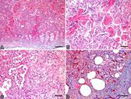
Histological features of idiopathic pulmonary fibrosis in felines. (A) Pulmonary parenchyma showing marked proliferation of spindle cells, especially in the subpleural region, associated with smooth muscle hypertrophy and reactive mesothelial cells. HE, bar = 250µm. (B) Marked hypertrophy of the terminal bronchiolar musculature and marked multifocal proliferation of type II pneumocytes. HE, bar = 130µm. (C) Marked multifocal proliferation of type II pneumocytes, infiltration of macrophages with a broad, sometimes foamy, cytoplasm and cellular debris within the alveoli, as well as proliferation of fibrous connective tissue. HE, bar = 130µm. (D) Pulmonary parenchyma showing a marked proliferation of fibrous connective tissue, distributed in a multifocal-to-coalescing form. MT, bar = 500µm.
On IHC, the proliferative spindle cells were remarkably reactive for vimentin (Fig.3A) and actin, which was also observed in the hyperplastic smooth muscle (Fig.3B). On IHC for pancytokeratin, there was an intense proliferation of type II pneumocytes around the alveoli (Fig.3C), and on IHC for lysozyme, an accumulation of alveolar macrophages was observed (Fig.3D).

Immunohistochemical evaluation of idiopathic pulmonary fibrosis in felines. (A) Intense intracytoplasmic diffuse staining of mesenchymal cells for vimentin. IHC and DAB, bar = 210µm. (B) Moderate and diffuse intracytoplasmic staining for smooth muscle actin. IHC and DAB, bar = 120µm. (C) Proliferation of type II pneumocytes around heavily immunoreactive alveoli for pancytokeratin. IHC and DAB, bar = 100µm. (D) Alveolar macrophages reactive for lysozyme. IHC and DAB, bar = 60µm.
There was no growth on bacteriological or mycological cultures, and immunohistochemical evaluations for the presence of viral etiological agents (FIV, FeLV, FCoV, FCV and FHV-1) was also negative.
Discussion
The diagnosis of idiopathic pulmonary fibrosis in the patients in this study was based on clinical, radiographic, pathological and immunohistochemical findings. All three cats were females ranging in age from 10 to 14 years old, which is the common age group of cats affected by this disease (Cohn et al. 2004Cohn L.A., Norris C.R., Hawkins E.C., Dye J.A., Johnson C.A. & Williams K.J. 2004. Identification and characterization of an idiopathic pulmonary fibrosis-Like condition in cats. J. Vet. Intern. Med. 18(5):632-641. <PMid:15515577>). As in humans, cats with pulmonary fibrosis are older, reflecting the insidious pathogenesis of the disease (Cohn et al. 2004Cohn L.A., Norris C.R., Hawkins E.C., Dye J.A., Johnson C.A. & Williams K.J. 2004. Identification and characterization of an idiopathic pulmonary fibrosis-Like condition in cats. J. Vet. Intern. Med. 18(5):632-641. <PMid:15515577>). Because the number of included cats was small, it was not possible to determine sex or breed predilection; however, the three patients in this study were females, as previously described in several other studies (Le Boedec et al. 2014Le Boedec K., Roady P.J. & O’Brien R.T. 2014. A case of atypical diffuse feline fibrotic lung disease. J. Feline Med. Surg. 16(10):858-863. <http://dx.doi.org/10.1177/1098612X14527963> <PMid:24650951>
https://doi.org/10.1177/1098612X14527963...
, Evola et al. 2014Evola M.G., Edmondson E.F., Reichle J.K., Biller D.S., Mitchell C.W. & Valdés-Martínez A. 2014. Radiographic and histopathologyc characteristics of pulmonary fibrosis in nine cats. Vet. Radiol. Ultrasound 55(2):133-140. <http://dx.doi.org/10.1111/vru.12106> <PMid:24103063>
https://doi.org/10.1111/vru.12106...
, Pereira et al. 2016Pereira C.O., Costa F.V.A., Gomes D.C., Kasper P.N., Vieira A.B. & Driemeier D. 2016. Dispneia expiratória restritiva em um gato com fibrose pulmonar idiopática: relato de caso. Arq. Bras. Med. Vet. Zootec. 69(3):593-596. <http://dx.doi.org/10.1590/1678-4162-8415>
https://doi.org/10.1590/1678-4162-8415...
).
These cats had similar clinical signs, characterized by anorexia, lethargy and respiratory distress with acute progression, similar to the previously reported clinical signs (Cohn et al. 2004Cohn L.A., Norris C.R., Hawkins E.C., Dye J.A., Johnson C.A. & Williams K.J. 2004. Identification and characterization of an idiopathic pulmonary fibrosis-Like condition in cats. J. Vet. Intern. Med. 18(5):632-641. <PMid:15515577>). Although this disease is characterized as a chronic condition, the clinical course of patients with idiopathic pulmonary fibrosis may be short due to the ability of these animals to mask pulmonary clinical signs (Cohn et al. 2004Cohn L.A., Norris C.R., Hawkins E.C., Dye J.A., Johnson C.A. & Williams K.J. 2004. Identification and characterization of an idiopathic pulmonary fibrosis-Like condition in cats. J. Vet. Intern. Med. 18(5):632-641. <PMid:15515577>). The radiographic changes were similar in the cats in this study, with adiffuse increase in radiopacity lung fields and a mixed bronchointerstitial pattern progressing to an alveolar pattern; however, some authors have stated that diffuse and heterogeneous lesions pattern are atypical in cats with fibrosis (Le Boedec et al. 2014Le Boedec K., Roady P.J. & O’Brien R.T. 2014. A case of atypical diffuse feline fibrotic lung disease. J. Feline Med. Surg. 16(10):858-863. <http://dx.doi.org/10.1177/1098612X14527963> <PMid:24650951>
https://doi.org/10.1177/1098612X14527963...
). However, the imaging features of this disease are variable (Cohn et al. 2004Cohn L.A., Norris C.R., Hawkins E.C., Dye J.A., Johnson C.A. & Williams K.J. 2004. Identification and characterization of an idiopathic pulmonary fibrosis-Like condition in cats. J. Vet. Intern. Med. 18(5):632-641. <PMid:15515577>, Evola et al. 2014Evola M.G., Edmondson E.F., Reichle J.K., Biller D.S., Mitchell C.W. & Valdés-Martínez A. 2014. Radiographic and histopathologyc characteristics of pulmonary fibrosis in nine cats. Vet. Radiol. Ultrasound 55(2):133-140. <http://dx.doi.org/10.1111/vru.12106> <PMid:24103063>
https://doi.org/10.1111/vru.12106...
) and may include a predominantly diffuse bronchointerstitial to alveolar pattern, as seen in this study. Pleural effusion, pulmonary nodules and mineralization are rarely observed (Evola et al. 2014Evola M.G., Edmondson E.F., Reichle J.K., Biller D.S., Mitchell C.W. & Valdés-Martínez A. 2014. Radiographic and histopathologyc characteristics of pulmonary fibrosis in nine cats. Vet. Radiol. Ultrasound 55(2):133-140. <http://dx.doi.org/10.1111/vru.12106> <PMid:24103063>
https://doi.org/10.1111/vru.12106...
); however, in the present study, two cats had mild to moderate pleural effusion. In terms of blood test results, there are no significant hematological alterations in both cats and humans (Cohn et al. 2004Cohn L.A., Norris C.R., Hawkins E.C., Dye J.A., Johnson C.A. & Williams K.J. 2004. Identification and characterization of an idiopathic pulmonary fibrosis-Like condition in cats. J. Vet. Intern. Med. 18(5):632-641. <PMid:15515577>).
The clinical presentation of this disease is similar to other pulmonary diseases in felines and due to the variation in radiographic findings and the absence of hematological and biochemical alterations, the clinical diagnosis is challenging (Cohn et al. 2004Cohn L.A., Norris C.R., Hawkins E.C., Dye J.A., Johnson C.A. & Williams K.J. 2004. Identification and characterization of an idiopathic pulmonary fibrosis-Like condition in cats. J. Vet. Intern. Med. 18(5):632-641. <PMid:15515577>). Procedures, such as bronchoscopy, were not useful for diagnosis in a previous study (Cohn et al. 2004Cohn L.A., Norris C.R., Hawkins E.C., Dye J.A., Johnson C.A. & Williams K.J. 2004. Identification and characterization of an idiopathic pulmonary fibrosis-Like condition in cats. J. Vet. Intern. Med. 18(5):632-641. <PMid:15515577>). Pulmonary aspiration may result in low cellularity samples because fibroblasts are juxtaposed cells, as observed in Cat 1. To achieve a definitive diagnosis histopathological evaluation of the lung is necessary (Cohn et al. 2004Cohn L.A., Norris C.R., Hawkins E.C., Dye J.A., Johnson C.A. & Williams K.J. 2004. Identification and characterization of an idiopathic pulmonary fibrosis-Like condition in cats. J. Vet. Intern. Med. 18(5):632-641. <PMid:15515577>, Pereira et al. 2016Pereira C.O., Costa F.V.A., Gomes D.C., Kasper P.N., Vieira A.B. & Driemeier D. 2016. Dispneia expiratória restritiva em um gato com fibrose pulmonar idiopática: relato de caso. Arq. Bras. Med. Vet. Zootec. 69(3):593-596. <http://dx.doi.org/10.1590/1678-4162-8415>
https://doi.org/10.1590/1678-4162-8415...
). However, because the treatment of the disease is limited, patients usually die after such an invasive diagnostic procedure, as occurred in two patients in this study (Cohn et al. 2004Cohn L.A., Norris C.R., Hawkins E.C., Dye J.A., Johnson C.A. & Williams K.J. 2004. Identification and characterization of an idiopathic pulmonary fibrosis-Like condition in cats. J. Vet. Intern. Med. 18(5):632-641. <PMid:15515577>, Le Boedec et al. 2014Le Boedec K., Roady P.J. & O’Brien R.T. 2014. A case of atypical diffuse feline fibrotic lung disease. J. Feline Med. Surg. 16(10):858-863. <http://dx.doi.org/10.1177/1098612X14527963> <PMid:24650951>
https://doi.org/10.1177/1098612X14527963...
). Oxygen therapy, bronchodilators, antimicrobials, diuretics and immunosuppressive drugs are used with no success or with a very short therapeutic response. Usually, the disease progresses very fast, leading to worsening respiratory conditions despite therapeutic attempts, as in the cases reported (Cohn et al. 2004Cohn L.A., Norris C.R., Hawkins E.C., Dye J.A., Johnson C.A. & Williams K.J. 2004. Identification and characterization of an idiopathic pulmonary fibrosis-Like condition in cats. J. Vet. Intern. Med. 18(5):632-641. <PMid:15515577>, Evola et al. 2014Evola M.G., Edmondson E.F., Reichle J.K., Biller D.S., Mitchell C.W. & Valdés-Martínez A. 2014. Radiographic and histopathologyc characteristics of pulmonary fibrosis in nine cats. Vet. Radiol. Ultrasound 55(2):133-140. <http://dx.doi.org/10.1111/vru.12106> <PMid:24103063>
https://doi.org/10.1111/vru.12106...
).
The cats in this study were domesticated and healthy; they did not have a historic of previous pulmonary disease. Based on IHC examinations, it was confirmed that these cats were negative for FIV, FeLV, FHV-1, FCV and FCoV, and there was no significant growth on bacteriological or mycological cultures, excluding the possibility of these infectious agents in the disease. As in humans, the pathogenesis of the disease in domestic felines has not been well elucidated. It has been suggested that, in humans, the onset of the condition is associated with pulmonary injury (Gross & Hunninghake 2001Gross T.J. & Hunninghake G.W. 2001. Idiopathic pulmonary fibrosis. Med. Prog. 345(7):517-525. <PMid:11519507>). Latent viral infection, particularly herpes viral infections, also has been reported as possible causes of pulmonary fibrosis (Gross & Hunninghake 2001Gross T.J. & Hunninghake G.W. 2001. Idiopathic pulmonary fibrosis. Med. Prog. 345(7):517-525. <PMid:11519507>). The same has been suggested for cats; however, the animals in this study were negative for the FHV-1 infection and were not previously diagnosed with any pulmonary parenchymal diseases that could have caused injury, such as feline asthma, pneumonia or neoplasia. Moreover, chemotherapy with nitrosourea for the treatment of intestinal lymphoma in felines has also been indicated as a cause of pulmonary fibrosis (Skorupski et al. 2008Skorupski K.A., Durham A.C., Duda L. & Sorenmo K.U. 2008. Pulmonary fibrosis after high cumulative dose nitrosourea chemotherapy in a cat. Vet. Comp. Oncol. 6(2):120-125. <http://dx.doi.org/10.1111/j.1476-5829.2007.00148.x> <PMid:19178671>
https://doi.org/10.1111/j.1476-5829.2007...
); however, none of the cats in this study had been treated with this medication. The microscopic and structural characteristics of type II pneumocytes observed in cats with the condition are similar to the familial form of this disease in humans, which occurs because of a defect in the gene encoding the surfactant protein C, suggesting a genetic component to the development of this disease (Van Moorsel et al. 2010Van Moorsel C.H., Van Oosterhout M.F., Barlo N.P., De Jong P.A., Van der Vis J.J., Ruven H.J., Van Es H.W., Van den Bosch J.M. & Grutters J.C. 2010. Surfactant protein C mutations are the basis of a significant portion of adult familial pulmonary fibrosis in a dutch cohort. Am. J. Respiratory Crit. Care Med. 182(11):1419-1425. <http://dx.doi.org/10.1164/rccm.200906-0953OC> <PMid:20656946>
https://doi.org/10.1164/rccm.200906-0953...
).
Pulmonary fibrosis may be observed in others domestic animals, such as in West Highland White Terriers dogs. These may present with a chronic progressive interstitial fibrosing lung disease of unknown etiology, with poor prognosis and no response to treatment (Heikkilä et al. 2011Heikkilä H.P., Lappalainen A.K., Day M.J., Clercx C. & Rajamäki M.M. 2011. Clinical, bronchoscopic, histopathologic, diagnostic imaging, and arterial oxygenation findings in West Highland white terriers with idiopathic pulmonary fibrosis. J. Vet. Intern. Med. 25(3):433-439. <http://dx.doi.org/10.1111/j.1939-1676.2011.0694.x> <PMid:21366693>
https://doi.org/10.1111/j.1939-1676.2011...
). The main finding of this condition is a diffuse interstitial fibrosis of the lung (Heikkilä et al. 2011Heikkilä H.P., Lappalainen A.K., Day M.J., Clercx C. & Rajamäki M.M. 2011. Clinical, bronchoscopic, histopathologic, diagnostic imaging, and arterial oxygenation findings in West Highland white terriers with idiopathic pulmonary fibrosis. J. Vet. Intern. Med. 25(3):433-439. <http://dx.doi.org/10.1111/j.1939-1676.2011.0694.x> <PMid:21366693>
https://doi.org/10.1111/j.1939-1676.2011...
), which is similar to that seen in cats affected by the same condition. Moreover, pulmonary fibrosis may be caused by specific etiological agents, such as the equine herpesvirus-5 (Panziera et al. 2014Panziera W., Giaretta P.R., Galiza G.J.N., Lim A., Bolin S.R., Borges C.H., Fighera R.A. & Barros C.S.L. 2014. Equine multinodular pulmonary fibrosis associated with equine herpesvirus 5 in a Horse in Brazil. Braz. J. Vet. Pathol. 7(1):17-20.), popularly called equine multinodular pulmonary fibrosis, that is a chronic infectious disease characterized by interstitial fibrosis of the lung in horses (Williams et al. 2007Williams K.J., Maes R., Del Piero F., Lim A., Wise A., Bolin D.C., Caswell J., Jackson C., Robinson N.E., Derksen F., Scott M.A., Uhal B.D., Li X., Youssef S.A. & Bolin S.R. 2007. Equine multinodular pulmonary fibrosis: a newly recognized herpesvirus-associated fibrotic lung disease. Vet. Pathol. 44(6):849-862. <http://dx.doi.org/10.1354/vp.44-6-849> <PMid:18039898>
https://doi.org/10.1354/vp.44-6-849...
). In humans, it is known that the inhalation of silica and asbestos may result in the development of fibrotic nodules (Oberdorster 1996Oberdorster G. 1996. Significance of particle parametersin the evaluation of exposure-dose-response relationships of inhaled particles. Inhalation Toxicol. 8(Suppl.):73-89. <PMid:11542496>). Interstitial fibrosis with concentric collagen deposition may occur in the lung of animals subsequently to chronic pulmonary hypertension, a condition caused by increased pulmonary vascular resistance or venous congestion (Caswell & Williams 2016Caswell J.L. & Williams K.J. 2016. Respiratory system, p.509-512. In: Maxie M.G. (Ed), Jubb, Kennedy, and Palmer’s Pathology of Domestic Animals. Vol.3. 6th ed. Elsevier, St Louis. <http://dx.doi.org/10.1016/B978-0-7020-5318-4.00011-5>.
https://doi.org/10.1016/B978-0-7020-5318...
). Moreover, additional histological changes of chronic pulmonary hypertension include intimal and medial hypertrophy in the pulmonary arterioles, as well as alveolar hemosiderophages (Caswell & Williams 2016Caswell J.L. & Williams K.J. 2016. Respiratory system, p.509-512. In: Maxie M.G. (Ed), Jubb, Kennedy, and Palmer’s Pathology of Domestic Animals. Vol.3. 6th ed. Elsevier, St Louis. <http://dx.doi.org/10.1016/B978-0-7020-5318-4.00011-5>.
https://doi.org/10.1016/B978-0-7020-5318...
).
Macroscopically, the lungs were firm with diffusely distributed lesions in the pulmonary parenchyma (Cohn et al. 2004Cohn L.A., Norris C.R., Hawkins E.C., Dye J.A., Johnson C.A. & Williams K.J. 2004. Identification and characterization of an idiopathic pulmonary fibrosis-Like condition in cats. J. Vet. Intern. Med. 18(5):632-641. <PMid:15515577>). The lesions were whitish and interspersed with areas of reddish color (mottled appearance). Other diseases may have similar appearance, such as chronic fungal or bacterial pneumonia and neoplasia (Le Boedec et al. 2014Le Boedec K., Roady P.J. & O’Brien R.T. 2014. A case of atypical diffuse feline fibrotic lung disease. J. Feline Med. Surg. 16(10):858-863. <http://dx.doi.org/10.1177/1098612X14527963> <PMid:24650951>
https://doi.org/10.1177/1098612X14527963...
). There is a known association between pulmonary fibrosis and pulmonary carcinoma (Aubry et al. 2002Aubry M.-C., Myers J.L., Douglas W.W., Tazelaar H.D., Stephens T.L.W., Hartman T.E., Deschamps C. & Pankratz V.S. 2002. Primary pulmonary carcinoma in patients with idiopathic pulmonary fibrosis. Mayo Clin. Proc. 77(8):763-770. <http://dx.doi.org/10.4065/77.8.763> <PMid:12173712>
https://doi.org/10.4065/77.8.763...
). There are several theories that have explained this association, including the progression of epithelial hyperplasia to neoplasia and the induction of carcinogenesis by chronic inflammation (Bouros et al. 2002Bouros D., Hatzakis K., Labrakis H. & Zeibecoglou K. 2002. Association of malignancy with diseases causing interstitial pulmonary changes. Chest J. 121(4):1278-1289. <http://dx.doi.org/10.1378/chest.121.4.1278> <PMid:11948064>
https://doi.org/10.1378/chest.121.4.1278...
).
The main microscopic lesion seen in cats with this disease is pulmonary fibrosis, as well as smooth muscle hyperplasia and type II pneumocyte proliferation (Cohn et al. 2004Cohn L.A., Norris C.R., Hawkins E.C., Dye J.A., Johnson C.A. & Williams K.J. 2004. Identification and characterization of an idiopathic pulmonary fibrosis-Like condition in cats. J. Vet. Intern. Med. 18(5):632-641. <PMid:15515577>). In all cases in this work, there was intense IHC staining for smooth muscle actin in proliferative spindle cells, suggesting the presence of myofibroblasts in the lesion. In most organs, tissue injury activates local fibrocytes that differentiate into contractile myofibroblasts, characterized by the expression of smooth muscle actin (Masseno et al. 2010Masseno A.P.B., Porto C.D., Nunes L.C., Sequeira J.L. & Alvarenga M.A. 2010. Miofibroblastos: revisão de literatura. Vet. Zootec. 17(2):177-190.). IHC for pancytokeratin showed a marked proliferation of type II pneumocytes around the alveoli. It is known that type II pneumocytes proliferate in order to repair injured tissue. Pneumocytes hinder gas exchange because these cells are cuboidal. This cuboidal form may no longer be observed on microscopy five to seven days after a mild injury, but it can remain present for a long period, in cases in which the injurious stimulus continues or in the presence of interstitial fibrosis (Caswell & Williams 2016Caswell J.L. & Williams K.J. 2016. Respiratory system, p.509-512. In: Maxie M.G. (Ed), Jubb, Kennedy, and Palmer’s Pathology of Domestic Animals. Vol.3. 6th ed. Elsevier, St Louis. <http://dx.doi.org/10.1016/B978-0-7020-5318-4.00011-5>.
https://doi.org/10.1016/B978-0-7020-5318...
). All of these findings suggest that pulmonary fibrosis in cats may be related to scarring from lung injury, whether the injury is ongoing or not. Although it is a routine in cat clinics to care for dyspneic cats, patients often die acutely without a diagnosis. Thus, it is necessary to consider pulmonary fibrosis as a differential diagnosis in cats with dyspnea, to increase the understanding of the pathogenesis of this disease and to determine an adequate prognosis and effective therapy in the future.
Conclusions
The animals were domesticated females ranging in age from 10 to 14 years old. The main clinical signs observed were mixed restrictive dyspnea, mild dehydration, and anorexia, and the clinical course lasted less than one month. The main radiographic findings were a mixed bronchointerstitial to alveolar pattern and a moderate diffuse increase in radiopacity in the lung fields.
Macroscopically, the lungs were firm with whitish areas and a slightly nodular appearance on the pleural surface, and in all cases, star-shaped scarring lesions with parenchymal retraction were present. Microscopically, the main findings were pulmonary fibrosis, type II pneumocyte proliferation, and hypertrophy or hyperplasia of smooth muscle tissue. On IHC, it was confirmed that the cats were negative for the main viral agents of the species.
References
- Aubry M.-C., Myers J.L., Douglas W.W., Tazelaar H.D., Stephens T.L.W., Hartman T.E., Deschamps C. & Pankratz V.S. 2002. Primary pulmonary carcinoma in patients with idiopathic pulmonary fibrosis. Mayo Clin. Proc. 77(8):763-770. <http://dx.doi.org/10.4065/77.8.763> <PMid:12173712>
» https://doi.org/10.4065/77.8.763 - Bouros D., Hatzakis K., Labrakis H. & Zeibecoglou K. 2002. Association of malignancy with diseases causing interstitial pulmonary changes. Chest J. 121(4):1278-1289. <http://dx.doi.org/10.1378/chest.121.4.1278> <PMid:11948064>
» https://doi.org/10.1378/chest.121.4.1278 - Caswell J.L. & Williams K.J. 2016. Respiratory system, p.509-512. In: Maxie M.G. (Ed), Jubb, Kennedy, and Palmer’s Pathology of Domestic Animals. Vol.3. 6th ed. Elsevier, St Louis. <http://dx.doi.org/10.1016/B978-0-7020-5318-4.00011-5>.
» https://doi.org/10.1016/B978-0-7020-5318-4.00011-5 - Cohn L.A., Norris C.R., Hawkins E.C., Dye J.A., Johnson C.A. & Williams K.J. 2004. Identification and characterization of an idiopathic pulmonary fibrosis-Like condition in cats. J. Vet. Intern. Med. 18(5):632-641. <PMid:15515577>
- Cushley M.J., Davison A.G. & Dubois R.M. 1999. The diagnosis, assessment and treatment of diffuse parenchymal lung disease in adults. Thorax 54(1):1-28. <PMid:10343621>
- Evola M.G., Edmondson E.F., Reichle J.K., Biller D.S., Mitchell C.W. & Valdés-Martínez A. 2014. Radiographic and histopathologyc characteristics of pulmonary fibrosis in nine cats. Vet. Radiol. Ultrasound 55(2):133-140. <http://dx.doi.org/10.1111/vru.12106> <PMid:24103063>
» https://doi.org/10.1111/vru.12106 - Gross T.J. & Hunninghake G.W. 2001. Idiopathic pulmonary fibrosis. Med. Prog. 345(7):517-525. <PMid:11519507>
- Heikkilä H.P., Lappalainen A.K., Day M.J., Clercx C. & Rajamäki M.M. 2011. Clinical, bronchoscopic, histopathologic, diagnostic imaging, and arterial oxygenation findings in West Highland white terriers with idiopathic pulmonary fibrosis. J. Vet. Intern. Med. 25(3):433-439. <http://dx.doi.org/10.1111/j.1939-1676.2011.0694.x> <PMid:21366693>
» https://doi.org/10.1111/j.1939-1676.2011.0694.x - Le Boedec K., Roady P.J. & O’Brien R.T. 2014. A case of atypical diffuse feline fibrotic lung disease. J. Feline Med. Surg. 16(10):858-863. <http://dx.doi.org/10.1177/1098612X14527963> <PMid:24650951>
» https://doi.org/10.1177/1098612X14527963 - Masseno A.P.B., Porto C.D., Nunes L.C., Sequeira J.L. & Alvarenga M.A. 2010. Miofibroblastos: revisão de literatura. Vet. Zootec. 17(2):177-190.
- McElroy D.A. 1992. Connective tissue, p.132. In: Prophet E.B., Mills B., Arrington J.B. & Sobin L.H. (Eds), Laboratory Methods in Histotechnology. Armed Forces Institute of Pathology, American Registry of Pathology, Washington.
- Oberdorster G. 1996. Significance of particle parametersin the evaluation of exposure-dose-response relationships of inhaled particles. Inhalation Toxicol. 8(Suppl.):73-89. <PMid:11542496>
- Panziera W., Giaretta P.R., Galiza G.J.N., Lim A., Bolin S.R., Borges C.H., Fighera R.A. & Barros C.S.L. 2014. Equine multinodular pulmonary fibrosis associated with equine herpesvirus 5 in a Horse in Brazil. Braz. J. Vet. Pathol. 7(1):17-20.
- Pereira C.O., Costa F.V.A., Gomes D.C., Kasper P.N., Vieira A.B. & Driemeier D. 2016. Dispneia expiratória restritiva em um gato com fibrose pulmonar idiopática: relato de caso. Arq. Bras. Med. Vet. Zootec. 69(3):593-596. <http://dx.doi.org/10.1590/1678-4162-8415>
» https://doi.org/10.1590/1678-4162-8415 - Rolim V.M., Casagrande R.A., Wouters A.T., Driemeier D. & Pavarini S.P. 2016. Myocarditis caused by feline immunodeficiency virus in five cats with hypertrophic cardiomyopathy. J. Comp. Path. 154(1):3-8. <http://dx.doi.org/10.1016/j.jcpa.2015.10.180> <PMid:26797583>
» https://doi.org/10.1016/j.jcpa.2015.10.180 - Selman M., Morrison L.D. & Noble P.W. 2010. Idiopathic interstitial pneumonias, p.1356-1397. In: Mason R.J., Broaddus V.C., Martin T.R., King T., Schraufnagel D., Murray J. & Nadel J. (Eds), Murray and Nadel’s Textbook of Respiratory Medicine. 5th ed. Saunders Elsevier, Philadelphia. <http://dx.doi.org/10.1016/B978-1-4160-4710-0.00057-2>.
» https://doi.org/10.1016/B978-1-4160-4710-0.00057-2 - Skorupski K.A., Durham A.C., Duda L. & Sorenmo K.U. 2008. Pulmonary fibrosis after high cumulative dose nitrosourea chemotherapy in a cat. Vet. Comp. Oncol. 6(2):120-125. <http://dx.doi.org/10.1111/j.1476-5829.2007.00148.x> <PMid:19178671>
» https://doi.org/10.1111/j.1476-5829.2007.00148.x - Thomas A.Q., Lane K., Phillips 3rd J., Prince M., Markin C., Speer M., Schwartz D.A., Gaddipati R., Marney A., Johnson J., Roberts R., Haines J., Stahlman M. & Loyd J.E. 2002. Heterozygosity for a surfactant protein c gene mutation associated with usual interstitial pneumonitis and cellular nonspecific interstitial pneumonitis in one kindred. Am. J. Respiratory Crit. Care Med. 165(9):1322-1328. <http://dx.doi.org/10.1164/rccm.200112-123OC> <PMid:11991887>
» https://doi.org/10.1164/rccm.200112-123OC - Van Moorsel C.H., Van Oosterhout M.F., Barlo N.P., De Jong P.A., Van der Vis J.J., Ruven H.J., Van Es H.W., Van den Bosch J.M. & Grutters J.C. 2010. Surfactant protein C mutations are the basis of a significant portion of adult familial pulmonary fibrosis in a dutch cohort. Am. J. Respiratory Crit. Care Med. 182(11):1419-1425. <http://dx.doi.org/10.1164/rccm.200906-0953OC> <PMid:20656946>
» https://doi.org/10.1164/rccm.200906-0953OC - Williams K.J., Maes R., Del Piero F., Lim A., Wise A., Bolin D.C., Caswell J., Jackson C., Robinson N.E., Derksen F., Scott M.A., Uhal B.D., Li X., Youssef S.A. & Bolin S.R. 2007. Equine multinodular pulmonary fibrosis: a newly recognized herpesvirus-associated fibrotic lung disease. Vet. Pathol. 44(6):849-862. <http://dx.doi.org/10.1354/vp.44-6-849> <PMid:18039898>
» https://doi.org/10.1354/vp.44-6-849
Publication Dates
-
Publication in this collection
Feb 2019
History
-
Received
04 Sept 2018 -
Accepted
18 Sept 2018


