
ABSTRACT:
B-scan ultrasonography is an important diagnostic tool that allows characterization of internal organ anatomy and, when complemented by Doppler ultrasound, allows vascular hemodynamic assessment, increasing the diagnostic accuracy. Thus, the aim of the present study was the B-scan ultrasound characterization and measurement of the eyeball segments and assessment of the external ophthalmic artery by color and pulsed Doppler. Sixty eyeballs were assessed from 30 dogs of different breeds using an 8.5MHz microconvex transductor. First, biometry was performed by B-scan of the following segments: axial length (M1), anterior chamber depth (M2), lens thickness (M3), lens length (M4), vitreous chamber depth (M5), optical disc length (M6) and optic nerve diameter (M7). Colored Doppler identified the external ophthalmic article and pulsed Doppler assessed its flow, and the following were measured: systolic peak velocity (VPS), final diastolic velocity (VDF), resistivity index (IR) and pulse index (IP). No statistical difference was observed for the biometric values of the eye segments between the right and left eyes (p>0.05). The vitreous chamber depth (M5) was shown to be the biometric variable with greatest bilateral symmetry, varying from 0.79 to 0.87cm and 0.78 to 0.86cm for the right and left eye, respectively. The ophthalmic artery was visualized over the optic nerve towards the eyeball, with flow stained red. There was no significant statistical difference between the Doppler velocimetric values for the ophthalmic artery between the right and left eye of the animals assessed (p>0.05). The mean resistivity index (RI) showed average values equal to 0.63±0.03, bilaterally. The mean base velocity was 17.50cm/s and 18.18cm/s at the systolic peak and 6.21cm/s and 6.68cm/s at the end of the diastole, for the right and left eyes respectively. The anatomic, biometric and hemodynamic characterization using the ultrasound B-scan and the Doppler modalities permitted the ultrasonographic and Doppler velocimetric assessment of the eyeball components in dogs of different breeds, and it can be used in ophthalmic clinical routine to identify eye pathologies.
INDEX TERMS:
Biometry; B-mode; Doppler ultrasound; dogs; eyeball; Doppler velocimetry; hemodynamics; ophthalmology
RESUMO:
O exame ultrassonográfico modo-B é uma importante ferramenta diagnóstica que permite caracterizar a anatomia interna dos órgãos e, complementada pelo exame modo Doppler, possibilita realizar a avaliação hemodinâmica vascular, aumentando a acurácia diagnóstica. Desta forma, este trabalho teve como objetivo a caracterização ultrassonográfica e mensuração dos segmentos do bulbo ocular modo-B, assim como a avaliação da artéria oftálmica externa pelo Doppler colorido e pulsado. Foram avaliados 60 bulbos oculares de 30 cães de diferentes raças utilizando transdutor microconvexo de 8,5MHz. Inicialmente foi realizada biometria por meio do exame em modo-B dos seguintes seguimentos: comprimento axial (M1), profundidade da câmara anterior (M2), espessura da lente (M3), comprimento da lente (M4), profundidade da câmara vítrea (M5), comprimento do disco óptico (M6) e diâmetro do nervo óptico (M7). A artéria oftálmica externa foi identificada pelo Doppler colorido e seu fluxo foi avaliado por meio do Doppler pulsado, sendo mensurados: velocidade do pico sistólico (VPS), velocidade diastólica final (VDF), índice de resistividade (IR) e índice de pulsatilidade (IP). Não foi verificada diferença estatística para os valores biométricos dos seguimentos oculares entre os olhos direito e esquerdo (p>0,05). A profundidade da câmera vítrea (M5), mostrou-se a variável biométrica com maior simetria bilateral, variando entre 0,79 a 0,87cm e 0,78 a 0,86cm para o olho direito e esquerdo, respectivamente. A artéria oftálmica foi visibilizada sobre o nervo óptico em direção ao bulbo ocular, com fluxo marcado em vermelho. Não houve diferença estatística significativa entre os valores Dopplervelocimétricos da artéria oftálmica entre do olho direito e esquerdo dos animais avaliados (p>0.05). O índice de resistividade (IR) médio evidenciou valores médios sendo igual a 0,63±0,03, bilateralmente. A velocidade basal média foi 17,50cm/s e 18,18cm/s no pico sistólico e 6,21cm/s e 6,68cm/s no final da diástole, para os olhos direito e esquerdo, respectivamente. A caracterização anatômica, biométrica e hemodinâmica utilizando o exame ultrassonográfico modo-B e as modalidades do Doppler permitiram a avaliação ultrassonográfica e Dopplervelocimétrica dos componentes do bulbo ocular nos cães de diferentes raças, podendo ser utilizados na rotina clínica oftalmológica para identificação de patologias oculares.
TERMOS DE INDEXAÇÃO:
Biometria; ultrassonografia; modo-B; Doppler colorido ocular; cães hígidos; bulbo ocular; Dopplervelocimetria; hemodinâmica; oftalmologia; caninos
Introduction
Eye ultrasound examination in veterinary medicine is considered a recent technique, but it is easily applied, does not require sedation and has no noxious effects on the patient. The technological progress that resulted in better image obtaining permits precise assessment of the eyeball and orbit structure, contributing to better diagnosis and therapeutic conduct (Ferreira et al. 2013Ferreira M.A., Cardoso K.C.F., Dias F.G.G., Brunelli A.T.J. & Honsho C.S. 2013. Ultrassonografia ocular como complemento do exame oftalmológico. Enciclopedia Biosfera 9(17):2487-2502.). However, because the ultrasound examination has been applied more to diagnose abdominal alterations in veterinary medicine, this technique is dominated by few professionals (Costa et al. 2014Costa A.P.A., Silva G.A., Lima A.M.V., Laus J.L. & Borges N.C. 2014. Ultrassonografia ocular em cães. Enciclopedia Biosfera 10(18):2905-2920.).
The ophthalmic ultrasonographic B-scan is used to assess eye periocular and retrobulbar structures (Laus et al. 2003Laus J.S., Canola J.C., Mamede F.V., Almeida D.E., Godoy G.S., Oliveira C.J.B., Pontin K., Albuquerque S. & Alessi A.C. 2003. Orbital cellulites associated with Toxocara canis in a dog. Vet. Ophthalmol. 6(4):333-336. http://dx.doi.org/10.1111/j.1463-5224.2003.00304.x. PMid:14641832.
https://doi.org/10.1111/j.1463-5224.2003...
, Tovar et al. 2005Tovar M.C., Huguet E. & Gomezi M.A. 2005. Orbital cellulitis and intraocular abscess caused by migrating grass in a cat. Vet. Ophthalmol. 8(5):353-356. http://dx.doi.org/10.1111/j.1463-5224.2005.00411.x. PMid:16178847.
https://doi.org/10.1111/j.1463-5224.2005...
, Singh & Young 2006Singh R.P. & Young L.H. 2006. Diagnostic tests for posterior segment inflammation. Int. Ophthalmol. Clin. 46(2):195-208. http://dx.doi.org/10.1097/00004397-200604620-00016. PMid:16770163.
https://doi.org/10.1097/00004397-2006046...
); in neoplasm afflictions (Baptista et al. 2006Baptista C.S., Villagrasa M. & Marinho A.A. 2006. Standardised B-scan and A-scan echographic evaluation of the spontaneous anterior uveal melanomas in the dog. Vet. J. 171(2):322-330. http://dx.doi.org/10.1016/j.tvjl.2004.11.005. PMid:16490716.
https://doi.org/10.1016/j.tvjl.2004.11.0...
) and in biometry, assessing variations in size, shape and position of these structures, helping in determining the prosthesis size and intra-eye pre-surgery (Hijar 2008Hijar M.V. 2008. Oftalmologia dos animais de companhia. Ultrassonografia Ocul. 3(1):49-62.).
Recently in dogs, another modality of ultrasound examination, the Doppler, has permitted determination of eye blood flow speed patterns (Feliciano et al. 2013Feliciano M.A.R., Abrahim M.A., Peixoto R.V.R., Yasunaga K.L., Vicente W.R.R. & Galera P.D. 2013. Contribution of ocular B-mode and triplex Doppler in the evaluation of 10 Poodle dogs with cataracts. Arq. Bras. Med. Vet. Zootec. 65(2):359-363. http://dx.doi.org/10.1590/S0102-09352013000200009.
https://doi.org/10.1590/S0102-0935201300...
). Using the technique measurements are taken of the flow that results in information about peripheral vascular resistance. In the pulsed Doppler modality, it is further possible to present information regarding the expression of resistance to blood flow inside an artery, through the resistivity index and pulsatility indices (Novellas et al. 2007Novellas R., Espada Y. & De Gopegui R.R. 2007. Doppler ultrasonographic estimation of renal and ocular resistive and pulsatility indices in normal dogs and cats. Vet. Radiol. Ultrasound 48(1):69-73. http://dx.doi.org/10.1111/j.1740-8261.2007.00206.x. PMid:17236363.
https://doi.org/10.1111/j.1740-8261.2007...
).
Thus, the objective of the present study was the ultrasound B-scan characterization of the eyeball and determination of Doppler velocimetric values of the external ophthalmic artery in healthy dogs of different breeds. The results of this study may help to identify biometric and hemodynamic alterations in animals with eye diseases, and aggregate important information to the ophthalmic veterinary routine.
Materials and Methods
Experimental design. Thirty dogs were studied (three daschunds, four pinschers, 10 crossbreeds and 13 poodles), 15 males and 15 females, aged 1 to 5 years, with no eye or systemic alteration at the clinical examination, weighing from 2.5 to 28.5kg, from the clinical medical services of the University Veterinary Hospital at the Federal University of Piauí, Brazil (HVU/UFPI). All the animals were submitted to the bilateral eye examination. The protocols used in the present study were approved by the Committee of Ethics in Animal Experimentation CEEA/UFPI (no. 135/16).
B-scan assessment. The ultrasound assessment of the eye components was made with the help of a portable ultrasound apparatus, model Z6-Vet (Myndray®, Shenzhen, China), associated to an 8.5MHz microconvex transductor (transdutor model, Myndray®, Shenzhen, China). The same observer made all the sweepings and the ultrasonography apparatus was calibrated always in the same way regarding the positioning of the focus and the brightness and contrast intensity, to prevent intra-observer variation while performing the examination.
The animals were placed in sternal decubitus and contained manually. For standardization, the right eye was assessed first and then the left eye. A drop of anesthetic proximetacaine chlorohydrate-based eyedrops (Anestalcon®, Alcon, São Paulo) was placeded in both the eyes to desensitize the cornea, and then a layer of carboxymethyl cellulose gel (Carbogel®, Carbogel Indústria e Comércio Ltda, São Paulo, Brazil) was applied over the transductor that was placed directly on the center of the cornea.
Sweepings were made on the sagittal and dorsal planes, and the following measurements were obtained: M1 = distance between the cornea and the internal face of the retina-choroid-sclera complex (axial length), M2 = distance between the central point of the cornea and the central point of the anterior lens capsule (anterior chamber depth), M3 = distance between the anterior and posterior lens capsule (lens thickness), M4 = Distance between the side and mid poles of the lens (lens length), M5 = distance between the posterior lens capsule and the posterior eyeball wall (vitreous chamber depth), M6 = optical disc length, M7 = optic nerve diameter (Fig.1).

Ultrasound image in B-scan showing the eyeball segments measured in biometry: axial length (M1), anterior chamber depth (M2), lens thickness (M3), lens length (M4), vitreous chamber depth (M5), optical disc length (M6) and optic nerve diameter (M7).
Doppler ultrasonography assessment. A sagittal cut was made in the eyeball. The external ophthalmic artery was identified by colored Doppler in topography of the optical nerve, at 2mm from the eyeball posterior wall. An insonation angle of less than 60° was used and the flow means were determined using the pulsed Doppler mode guided by the colored Doppler (duplex mode). The means derived from the wave spectrum in pulsed Doppler (RI = resistivity index and PI = pulsatility index) were obtained after manual marking of the maximum systolic peak velocity (VPS) and the final diastolic velocity (VDF).
All the measurements in B-scan and Doppler were acquired in triplicate and the mean of the values obtained was used for the results.
At the end of the examination both the eyes were cleaned with a 0.9% sodium chloride solution to prevent possible irritative processes caused by the gel.
Statistical analysis. The data were submitted to the normality test (the Shapiro-Wilk and Kolmogorov-Smirnov test) and later the means were analyzed by the student t-test to interpret the parameters, considering a 5% confidence interval.
Results
B-scan ultrasound examination
The B-scan ultrasound examination was shown to be an easily applied technique, permitting the identification, assessment and measurement of all the components of the eyeball in the animals assessed.
The cornea was characterized by a hyperechogenic line. The anterior chamber was visualized as a space located between the cornea and the anterior lens face, filled with liquid with anechogenic characteristics (aqueous humor). The iris and the ciliary body, identified in the lens peripheral-equatorial region, were presented as a single structure in the sagittal cut that was triangular and echogenic in shape. The lens was characterized by a convex structure, formed by an anterior ecogenic face and another posterior, shortly after the anterior chamber, and were occupied internally by anechogenic content. The vitreous space, located between the lens posterior face and the posterior wall of the eyeball, was filled with anechogenic liquid (vitreous humor). The optical disc was identified on the posterior wall of the eyeball, slightly more echogenic than the wall and the optical nerve, caudal to the optical disc, and was shown as a funnel-shaped structure, hypoechogenic in relation to the adjacent retrobulbar tissue (Fig.1).
The biometric measurements of the eyeball components are presented in Table 1. Comparison of the means of the eye structures between the right and left eyes showed bilateral symmetry, and there was no significant statistical difference between the axial length (M1), anterior chamber depth (M2), lens thickness (M3), lens length (M4), vitreous chamber depth (M5), optical disc length (M6) and optic nerve diameter (M7) (p>0.05). All the biometric values presented very close values when both eyes were compared and the vitreous chamber depth (M5) was the biometric variable with greatest bilateral symmetry, ranging from 0.79 to 0.87cm and 0.78 to 0.86cm for the right and left eye, respectively.
Positive correlation was observed between the biometric measurements of the eyeball and the animal´s age (Table 2) and the correlation coefficients varied from 0.62 to 0.96. Strong dependence was observed in 78.57% of the observations.
Doppler mode ultrasonography
Doppler ultrasonography was shown to be an important tool in assessing the hemodynamic components of the eyeball and the retrobulbar structures, permitting the identification of the external ophthalmic artery, characterization of its path and obtaining the Doppler velocimetric indices, complementing the B-scan assessment.
The external ophthalmic artery was observed in both the eyes of all the animals in the study, with a path over the optic nerve in direction of the eyeball. It was characterized by the presence of intermediate resistivity parabolic laminar flow with high systolic peak and dycrotic trace (Fig.2).
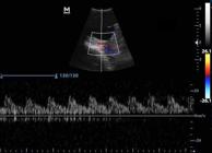
Triplex Doppler image of the external ophthalmic artery. Pattern of parabolic laminar flow with intermediate resistivity and presence of dicrotic trace.
Table 3 shows the Doppler velocimetric values of the external ophthalmic artery. The hemodynamic indices measured do not show statistical difference (p>0.05) when the right and left eyes are compared. The mean resistivity index (RI) showed mean values equal to 0.63±0.03, bilaterally. The mean base velocity was 17.50cm/s and 18.18cm/s at the systolic peak and 6.21cm/s and 6.6cm/s at the end of the diastole for the right and left eyes respectively.
There was partial and positive correlation between the animals´ age and the systolic peak velocity (SPV) and the resistivity index (RI) for both eyes, with values ranging from 0.80 to 0.89 (Table 4).
Discussion
The two-dimensional ultrasound examination used to assess the eyeball of the dogs in the present study allowed the evaluation of the internal eye anatomy, without iatrogenic injury to the cornea surface, as corroborated by Soares et al. (1998)Soares A.M.B., Laus J.L., Siqueira Y.H. & Marsillac P. 1998. Ultra-sonografia bidimensional em tempo real do bulbo ocular de cães (Canis familiaris, Linnaeus, 1758) com opacificação de meios transparentes. Emprego do transdutor mecânico setorial de 7,5 MHz com almofada de recuo. Ciência Rural 28(4):591-599. http://dx.doi.org/10.1590/S0103-84781998000400010.
https://doi.org/10.1590/S0103-8478199800...
and Gonçalves et al. (2009)Gonçalves G.F., Leme M.C., Romagnolli P., Eurides D. & Pippi N.L. 2009. Biometria ultrassonográfica bidimensional em tempo real de bulbo ocular de gatos domésticos. Ciênc. Anim. Bras. 10(3):829-834..
The transcorneal technique used in the present study permitted identification of the eyeball structures in all the animals. According to Hager et al. (1987)Hager D.A., Dziezyc J. & Millchamp N.J. 1987. Two-dimensional real-time ocular ultrasonography in the dog. Vet. Radiol. 28(2):60-65. http://dx.doi.org/10.1111/j.1740-8261.1987.tb01726.x.
https://doi.org/10.1111/j.1740-8261.1987...
and Hermsen (1984)Hermsen V. 1984. The use of ultrasound in the evaluation of diabetic vitreretinopathy. Int. Ophthalmol. Clin. 24(4):125-141. http://dx.doi.org/10.1097/00004397-198402440-00012. PMid:6389408.
https://doi.org/10.1097/00004397-1984024...
, this technique permits a more reliable examination of the eye elements, because the transpalpebral techniques result in loss of more than 50% given echo return. Only the cornea thickness measurement could not be assessed because of the low frequency of the transductor used (8.5MHz). Beserra et al. (2009)Beserra P.S., Sales G.A., Santana E.J.M., Miranda S.A., Brito A.B., Nickolak E. & Domingues S.F.S. 2009. Relação entre a biometria ultra-sonográfica em modo B do bulbo ocular e os diâmetros frontoocciptal e bizigomático em Canis familiaris. Pesq. Vet. Bras. 29(4):286-290. http://dx.doi.org/10.1590/S0100-736X2009000400002.
https://doi.org/10.1590/S0100-736X200900...
assessed dogs using a 5 to 8MHz frequency microconvex transductor and reported the same phenomenon. Ferreira et al. (2014)Ferreira M.A., Allemann N., Dias L.G.G.G. & Honsho C.S. 2014. Relação entre a biometria ultrassonográfica ocular e os parâmetros morfométricos do crânio, idade, peso e gênero em gatos domésticos. Pesq. Vet. Bras. 34(2):192-198. http://dx.doi.org/10.1590/S0100-736X2014000200016.
https://doi.org/10.1590/S0100-736X201400...
demonstrated the inefficiency of frequencies between 8 and 9MHz when measuring cornea thickness in adult cats. Transductors with frequency higher than 20MHz give greater image definition and penetration power (from 20 to 80μm), allowing the visualization and measurement of images of the anterior eye segment, including the cornea in dogs and cats (Bentley et al. 2003Bentley E., Miller P.E. & Diehl K.A. 2003. Use of high-resolution ultrasound as a diagnostic tool in veterinary ophthalmology. J. Am. Vet. Med. Assoc. 223(11):1617-1622. http://dx.doi.org/10.2460/javma.2003.223.1617. PMid:14664449.
https://doi.org/10.2460/javma.2003.223.1...
).
In veterinary medicine, initial studies on biometric parameters began with Schiffer et al. (1982)Schiffer S.P., Rantanen N.W., Leary C.A. & Bryan G.M. 1982. Biometric study of the canine eye, using A-mode ultrasonography. Am. J. Vet. Res. 43(5):826-830. PMid:7091846. in dogs using A-scan ultrasound. In wild animals, B-scan biometric assessments were carried out on koalas (Hirst et al. 1992Hirst L.W., Brown A.S., Kempster R. & Winney N. 1992. Ophthalmologic examination of the normal eye of the koala. J. Wildl. Dis. 28(3):419-423. http://dx.doi.org/10.7589/0090-3558-28.3.419. PMid:1512874.
https://doi.org/10.7589/0090-3558-28.3.4...
), alligators (Maia et al. 2003Maia F.B.N., Pigatto J.A.T., Guedes P.M. & Barros P.S.M. 2003. Ocular biomicroscopy (UBM) in Yacare Caiman (Caiman yacare). Vet. Ophthalmol. 6(4):351-366.), ferrets (Hernandez-Guerra et al. 2007Hernandez-Guerra A.M., Rodilla V. & López-Murcia M.M. 2007. Ocular biometry in the adult anesthetized ferret (Mustela putorius furo). Vet. Ophthalmol. 10(1):50-52. http://dx.doi.org/10.1111/j.1463-5224.2007.00500.x. PMid:17204128.
https://doi.org/10.1111/j.1463-5224.2007...
), elephants (Nunnery et al. 2008Nunnery C.M., Barrie K.P., Wiendner E.B., Gellatt-Nicholson K.J., Plummer C.E. & Brooks D.E. 2008. Ocular ultrasound findings in the Asian elephant Elephas maximus. Vet. Ophthalmol. 11(6):411-419.) and parrots (Lehmkuhl et al. 2010Lehmkuhl R.C., Almeida M.F., Mamprim M.J. & Vulcano L.C. 2010. B-mode ultrasonography iometry of the Amazon Parrot (Amazona aestiva) eye. Vet. Ophthalmol. 13(1):26-28. http://dx.doi.org/10.1111/j.1463-5224.2010.00797.x. PMid:20149172.
https://doi.org/10.1111/j.1463-5224.2010...
).
According to the statistical analysis, there was no evidence of difference (p>0.05) in the measurements of the eyeball segments between the right and left eyes (Table 1), that presented great homogeneity, as reported by Schiffer et al. (1982)Schiffer S.P., Rantanen N.W., Leary C.A. & Bryan G.M. 1982. Biometric study of the canine eye, using A-mode ultrasonography. Am. J. Vet. Res. 43(5):826-830. PMid:7091846., Gaiddon et al. (1991)Gaiddon J., Rosolen S.G., Steru L., Cook C.S. & Peiffer Junior R. 1991. Use of biometry and keratometry for determining optimal power for intraocular lens implants in dogs. Am. J. Vet. Res. 52(5):781-783. PMid:1854106., Williams (2004)Williams D.L. 2004. Lens morphometry determined by B-mode ultrasonography of the normal and cataractous canine lens. Vet. Ophthalmol. 7(2):91-95. http://dx.doi.org/10.1111/j.1463-5224.2004.04005.x. PMid:14982588.
https://doi.org/10.1111/j.1463-5224.2004...
, Boroffka et al. (2006)Boroffka S.A.E.B., Voorhout G., Verbruggen A.M. & Teske E. 2006. Intraobserver and interobserver repeatability of ocular biometric measurements obtained by means B-mode ultrasonography in dogs. Am. J. Vet. Res. 67(10):1743-1749. http://dx.doi.org/10.2460/ajvr.67.10.1743. PMid:17014326.
https://doi.org/10.2460/ajvr.67.10.1743....
, Tuntivanich et al. (2007)Tuntivanich N., Petersen-jones S.M., Steibel J.P., Johnson C. & Forcier J.Q. 2007. Postnatal development of canine axial globe length measured by B-scan ultrasonography. Vet. Ophthalmol. 10(1):2-5. http://dx.doi.org/10.1111/j.1463-5224.2007.00481.x. PMid:17204121.
https://doi.org/10.1111/j.1463-5224.2007...
and Beserra et al. (2009)Beserra P.S., Sales G.A., Santana E.J.M., Miranda S.A., Brito A.B., Nickolak E. & Domingues S.F.S. 2009. Relação entre a biometria ultra-sonográfica em modo B do bulbo ocular e os diâmetros frontoocciptal e bizigomático em Canis familiaris. Pesq. Vet. Bras. 29(4):286-290. http://dx.doi.org/10.1590/S0100-736X2009000400002.
https://doi.org/10.1590/S0100-736X200900...
.
The axial length was 1.75±0.10 and 1.73±0.11cm for the right and left eyes, respectively (Table 1). Gonçalves et al. (2000)Gonçalves G.F., Pippi N.L., Raiser A.G., Mazzanti A., Oliveira S.T., Neves J.P., Leotte A.M. & Hintz C.W. 2000. Biometria ultra-sonográfica bidimensional em tempo real do globo ocular de cães. Ciência Rural 30(3):417-420. http://dx.doi.org/10.1590/S0103-84782000000300007.
https://doi.org/10.1590/S0103-8478200000...
found values above those of the present study when they assessed crossbred dogs weighing between 5 and 12kg (1.88±0.8cm). Studies by Cottrill et al. (1989)Cottrill N.B., Banks W.J. & Pechman R.D. 1989. Ultrasonographic and biometric evalution of the eye and orbit of dogs. Am. J. Vet. Res. 50(6):898-903. PMid:2669574. compared the axial length of enucleated eyes of mesocephalic and dolicocephal dogs and reported that the values measured in mesocephalic dogs (1.99±1.2cm) were smaller than in dolicocephal dogs (2.12±1.3cm), suggesting that the longer the cranium, the longer will be the axial length found. In human beings, the variation in the axial length is approximately 0.5cm (François & Goes 1977François J. & Goes F. 1977. Ultrassonographic study of 100 emmetropic eyes. Ophthalmologica 175(6):321-327. http://dx.doi.org/10.1159/000308676. PMid:593658.
https://doi.org/10.1159/000308676....
), while in the present study, the axial length varied by about 0.1cm.
The anterior chamber depth ranged from 0.15 to 0.25cm for the right eye and from 0.13 to 0.25cm for the left eye (Table 1). According to Miller & Murphy (1995)Miller P.E. & Murphy C.J. 1995. Visionin Dogs. J. Am. Vet. Med. Assoc. 207(12):1623-1634. PMid:7493905. the anterior chamber depth is the measurement most affected by errors in two-dimensional mode biometry because the cornea is flattened by the transductor, leading to a decrease in its depth of approximately 0.14cm. In a study carried out with 30 crossbred dogs, significant statistical difference was observed between the right and left eyes (Gonçalves et al. 2000Gonçalves G.F., Pippi N.L., Raiser A.G., Mazzanti A., Oliveira S.T., Neves J.P., Leotte A.M. & Hintz C.W. 2000. Biometria ultra-sonográfica bidimensional em tempo real do globo ocular de cães. Ciência Rural 30(3):417-420. http://dx.doi.org/10.1590/S0103-84782000000300007.
https://doi.org/10.1590/S0103-8478200000...
). This difference can be explained by the use of a low frequency transductor (7.5 MHz) compared to the frequency used in the present research (8.5 MHz), leading to the formation of images with less resolution power, or even by the flattening phenomenon. Not using the standoff pad did not interfere in identifying and measuring the anterior chamber depth, because the gel used formed a thick layer between the transductor and the cornea, permitting its complete analysis, as proposed by Cottrill et al. (1989)Cottrill N.B., Banks W.J. & Pechman R.D. 1989. Ultrasonographic and biometric evalution of the eye and orbit of dogs. Am. J. Vet. Res. 50(6):898-903. PMid:2669574.. Furthermore, all the images were obtained by the same apparatus and taken by a single, experienced examinationiner, according to recommendations by Findl et al. (2003)Findl O., Kriechbaum K., Sacu S., Kiss B., Polak K., Nepp J., Schild G., Rainer G., Maca S., Petternel V., Lackner B. & Drexler W. 2003. Influence of operator experience on the performance of ultrasound biometry compared to optical biometry before catarata surgery. J. Cataract Refract. Surg. 29(10):1950-1955. http://dx.doi.org/10.1016/S0886-3350(03)00243-8. PMid:14604716.
https://doi.org/10.1016/S0886-3350(03)00...
that were factors favorable to the biometric measurements.
The means for the lens thickness and length were 0.72±0.05cm for the right eye and 0.73±0.04cm for the left eye; and 1.22±0.10cm for the right eye and 1.22±0.11cm for the left eye, respectively, and the values were very close when both eyes were compared (Table 1). Silva et al. (2010)Silva M.L., Martins B.C., Ribeiro A.P., Souza A.L.G. & Laus J.L. 2010. A- and B-modes echobiometry in cataractous and noncataractous eyes of English Cocker Spaniel dogs. Arq. Bras. Med. Vet. Zootec. 62(5):1080-1085. http://dx.doi.org/10.1590/S0102-09352010000500009.
https://doi.org/10.1590/S0102-0935201000...
assessed seven dogs of the English Cocker Spaniel breed and observed mean lens thickness of 7.14±0.35mm for the right eye and 7.19±0.57mm for the left eye that presented very close values compared to the present study. In this same study, biometric assessment was also made of the eyeball of dogs with cataract and it was verified that there was no statistical difference between the animals in the control group and the group with cataracts.
The vitreous chamber depth varied from 0.79 to 0.87cm, 0.82cm mean (Table 1). The same measurement was reported by Cottrill et al. (1989)Cottrill N.B., Banks W.J. & Pechman R.D. 1989. Ultrasonographic and biometric evalution of the eye and orbit of dogs. Am. J. Vet. Res. 50(6):898-903. PMid:2669574. ranging from 0.9 to 1.0cm, mean 0.96±0.16cm, and higher than the values found in the present study. Gonçalves et al. (2000)Gonçalves G.F., Pippi N.L., Raiser A.G., Mazzanti A., Oliveira S.T., Neves J.P., Leotte A.M. & Hintz C.W. 2000. Biometria ultra-sonográfica bidimensional em tempo real do globo ocular de cães. Ciência Rural 30(3):417-420. http://dx.doi.org/10.1590/S0103-84782000000300007.
https://doi.org/10.1590/S0103-8478200000...
found mean values of 9.1±0.5mm for the right eye and 9.1±0.4mm for the left eye, when they studied crossbred dogs. This difference may be related to the large variation of breeds among the animals studied, because the stand and size of the animals may interfere in the size of the intraeye structures (Sampaio et al. 2002Sampaio G.R., Ranzani J.J.T. & Schellini S.A. 2002. Sexo, peso e conformação anatômica do olho sobre o cálculo de poder dióptrico de lentes intra-oculares no cão. Ciência Rural 32(2):263-268. http://dx.doi.org/10.1590/S0103-84782002000200013.
https://doi.org/10.1590/S0103-8478200200...
). Cottrill et al. (1989)Cottrill N.B., Banks W.J. & Pechman R.D. 1989. Ultrasonographic and biometric evalution of the eye and orbit of dogs. Am. J. Vet. Res. 50(6):898-903. PMid:2669574. further stated that dolichocephalic dogs present high values for vitreous chamber depth when compared to mesocephalic dogs. The vitreous chamber depth is an important measurement to assess the retina anatomic integrity in cases where the animal has some degree of anterior opacity in the eye, and ultrasound assessment is recommended of areas of displacement or in conditions of eyeball rupture (Gonçalves et al. 2000Gonçalves G.F., Pippi N.L., Raiser A.G., Mazzanti A., Oliveira S.T., Neves J.P., Leotte A.M. & Hintz C.W. 2000. Biometria ultra-sonográfica bidimensional em tempo real do globo ocular de cães. Ciência Rural 30(3):417-420. http://dx.doi.org/10.1590/S0103-84782000000300007.
https://doi.org/10.1590/S0103-8478200000...
).
The optic nerve measurements presented a mean of 0.41±0.08cm, and the optical disc 0.39±0.05cm, and there were no statistical differences between the right and left eyes for either structure. The B-scan ultrasound examination can show distension of the optic nerve sheath (Urbano et al. 2002Urbano A.P., Urbano A.P., Urbano I. & Kara-José N. 2002. Episclerite e esclerite. Arq. Bras. Oftalmol. 65(5):591-598. http://dx.doi.org/10.1590/S0004-27492002000500018.
https://doi.org/10.1590/S0004-2749200200...
).
There was positive correlation between the biometric values of the eyeball structures and the age of the dogs, that suggests that eye growth increases according to the age of the animal, that is, their growth. This finding was also reported by Tuntivanich et al. (2007)Tuntivanich N., Petersen-jones S.M., Steibel J.P., Johnson C. & Forcier J.Q. 2007. Postnatal development of canine axial globe length measured by B-scan ultrasonography. Vet. Ophthalmol. 10(1):2-5. http://dx.doi.org/10.1111/j.1463-5224.2007.00481.x. PMid:17204121.
https://doi.org/10.1111/j.1463-5224.2007...
. Boroffka (2005)Boroffka S.A.E.B. 2005. Ultrasonography evaluation of pre- and postnatal development of the eye in Beagles. Vet. Radiol. Ultrasound 46(1):72-79. http://dx.doi.org/10.1111/j.1740-8261.2005.00015.x. PMid:15693565.
https://doi.org/10.1111/j.1740-8261.2005...
assessed the intraeye structure development in four fetuses in prenatal care and in 11 puppies in postnatal care using B-scan ultrasonography and observed that both in the pre-and post natal care there was continuous growth of the eye depth, anterior chamber, lens and vitreous chamber.
Ultrasound with Doppler mapping is a technique used complementary to the B-scan and allows non-invasive study of local vascularization, identifying possible alterations of vessel caliber and pathway and determining quantitative blood flow and vascular resistance parameters (Carvalho et al. 2008Carvalho C.F., Chammas M.C. & Cerri G.G. 2008. Princípios físicos do Doppler em ultrassonografia: revisão bibliográfica. Ciência Rural 38(3):872-879. http://dx.doi.org/10.1590/S0103-84782008000300047.
https://doi.org/10.1590/S0103-8478200800...
).
The first studies on using the Doppler mode on ophthalmic vascularization in normal dogs were by Gelatt-Nicholson et al. (1999)Gelatt-Nicholson, Gelatt, MacKay, Brooks & Newell S.M. 1999. Doppler imaging of the ophthalmic vasculature of the normal dog: blood velocity measurements and reproducibility. Vet. Ophthalmol. 2(2):87-96. http://dx.doi.org/10.1046/j.1463-5224.1999.00062.x. PMid:11397248.
https://doi.org/10.1046/j.1463-5224.1999...
. Later studies were made on felines (Gonçalves et al. 2005Gonçalves G.F., Pippi N.L., Leme M.C., Custódio A.P., Fachin L., Lago E., Silva A.V. & Pachaly J.R. 2005. Fluxometria Eco-Power-Doppler da artéria oftálmica externa em gatos (Felis catus Linnaeus, 1758). Arq. Ciênc. Vet. Zool. 8(2):117-124.), rabbits (Liu et al. 2007Liu J.H.K., Li R., Nelson T.R. & Weinreb R. 2007. Resistance to blood flow in the rabbit ophthalmic artery after topical treatment with timolol. J. Ocul. Pharmacol. Ther. 23(2):103-109. http://dx.doi.org/10.1089/jop.2006.0098. PMid:17444797.
https://doi.org/10.1089/jop.2006.0098....
) and sheep (Gerometta et al. 2010Gerometta R., Alvarez L.J. & Candia O.A. 2010. Effects of sildenafil and tadalafil on intraocular pressure in sheep: implications for aqueous humor dynamics. Invest. Ophthalmol. Vis. Sci. 51(6):3139-3144. http://dx.doi.org/10.1167/iovs.09-4862. PMid:20089876.
https://doi.org/10.1167/iovs.09-4862....
).
The external ophthalmic artery presented a dorsal pathway to the optical nerve, with flow towards the eyeball, and was visualized with red stain by colored Doppler, that is, in the direction of the transductor, like that described by Gonçalves et al. (2005)Gonçalves G.F., Pippi N.L., Leme M.C., Custódio A.P., Fachin L., Lago E., Silva A.V. & Pachaly J.R. 2005. Fluxometria Eco-Power-Doppler da artéria oftálmica externa em gatos (Felis catus Linnaeus, 1758). Arq. Ciênc. Vet. Zool. 8(2):117-124.. Pulsed Doppler classified the flow as parabolic with intermediate resistivity with high systolic peak and dicrotic trace (Fig.2Bentley E., Miller P.E. & Diehl K.A. 2003. Use of high-resolution ultrasound as a diagnostic tool in veterinary ophthalmology. J. Am. Vet. Med. Assoc. 223(11):1617-1622. http://dx.doi.org/10.2460/javma.2003.223.1617. PMid:14664449.
https://doi.org/10.2460/javma.2003.223.1...
), like the assessment of the external ophthalmic artery in cats (Gonçalves et al. 2005Gonçalves G.F., Pippi N.L., Leme M.C., Custódio A.P., Fachin L., Lago E., Silva A.V. & Pachaly J.R. 2005. Fluxometria Eco-Power-Doppler da artéria oftálmica externa em gatos (Felis catus Linnaeus, 1758). Arq. Ciênc. Vet. Zool. 8(2):117-124.) and rabbits (Liu et al. 2007Liu J.H.K., Li R., Nelson T.R. & Weinreb R. 2007. Resistance to blood flow in the rabbit ophthalmic artery after topical treatment with timolol. J. Ocul. Pharmacol. Ther. 23(2):103-109. http://dx.doi.org/10.1089/jop.2006.0098. PMid:17444797.
https://doi.org/10.1089/jop.2006.0098....
). In human medicine, the ophthalmic artery flow in normal pregnant women is characterized by a mono-phase wave, with slow systolic ascension and discreetly rounded peak, followed by two small increases in flow during the diastole (dicrotic pattern) (Diniz et al. 2005Diniz A.L.D., Moron A.F., Santos M.C., Sass N. & Pires C.R. 2005. Dopplervelocimetria das artérias oftálmica e central da retina em gestantes normais. Revta Bras. Ginecol. Obstet. 27(4):168-173. http://dx.doi.org/10.1590/S0100-72032005000400002.
https://doi.org/10.1590/S0100-7203200500...
).
The external ophthalmic artery flow can be influenced during blood passage through its interior by the blood volume injected, vessel wall resistance and elasticity. Thus, it is expected that the blood flow velocity inside this artery is bigger during the systole and smaller in the diastole (Grosenbaugh & Muir 1998Grosenbaugh D.A. & Muir 3rd W.W. 1998. Blood pressure monitoring. Am. J. Vet. Res. 59(2):205-212. PMid:9492938., Gonçalves et al. 2005Gonçalves G.F., Pippi N.L., Leme M.C., Custódio A.P., Fachin L., Lago E., Silva A.V. & Pachaly J.R. 2005. Fluxometria Eco-Power-Doppler da artéria oftálmica externa em gatos (Felis catus Linnaeus, 1758). Arq. Ciênc. Vet. Zool. 8(2):117-124.). According to Carvalho et al. (2008)Carvalho C.F., Chammas M.C. & Cerri G.G. 2008. Princípios físicos do Doppler em ultrassonografia: revisão bibliográfica. Ciência Rural 38(3):872-879. http://dx.doi.org/10.1590/S0103-84782008000300047.
https://doi.org/10.1590/S0103-8478200800...
the recognition of characteristic patterns of pulsed wave spectrum to Doppler in each tissue allows identification and observation of pathological alterations.
The mean resistivity index both for the right external ophthalmic artery and for the left external ophthalmic artery was the same, 0.63±0.03 (Table 3), and was considered of intermediate resistivity. Similar values were found by Novellas et al. (2007)Novellas R., Espada Y. & De Gopegui R.R. 2007. Doppler ultrasonographic estimation of renal and ocular resistive and pulsatility indices in normal dogs and cats. Vet. Radiol. Ultrasound 48(1):69-73. http://dx.doi.org/10.1111/j.1740-8261.2007.00206.x. PMid:17236363.
https://doi.org/10.1111/j.1740-8261.2007...
who assessed healthy dogs of different breeds and found mean values of 0.63±0.06. Gelatt-Nicholson et al. (1999)Gelatt-Nicholson, Gelatt, MacKay, Brooks & Newell S.M. 1999. Doppler imaging of the ophthalmic vasculature of the normal dog: blood velocity measurements and reproducibility. Vet. Ophthalmol. 2(2):87-96. http://dx.doi.org/10.1046/j.1463-5224.1999.00062.x. PMid:11397248.
https://doi.org/10.1046/j.1463-5224.1999...
found values of 0.58±0.1 below those found in the present study, which may be related to the small number of the animals in the study. Feliciano et al. (2013)Feliciano M.A.R., Abrahim M.A., Peixoto R.V.R., Yasunaga K.L., Vicente W.R.R. & Galera P.D. 2013. Contribution of ocular B-mode and triplex Doppler in the evaluation of 10 Poodle dogs with cataracts. Arq. Bras. Med. Vet. Zootec. 65(2):359-363. http://dx.doi.org/10.1590/S0102-09352013000200009.
https://doi.org/10.1590/S0102-0935201300...
studied dogs with cataract and observed an increase in the resistance of the vascular bed of the ophthalmic artery, with means of 0.76±0.1 for the right eye and 0.72±0.09 for the left eye, and most of the animals presented turbulent arterial flow.
Lee et al. (2002)Lee H., Chang D., Lee Y., Eom K., Choi H., Seo K., Choi M. & Yoon J. 2002. Use of color Doppler imaging for determining the resistive index of the medial long posterior ciliary artery in clinically normal conscious dogs. Am. J. Vet. Res. 63(2):211-214. http://dx.doi.org/10.2460/ajvr.2002.63.211. PMid:11843120.
https://doi.org/10.2460/ajvr.2002.63.211...
reported that the Doppler ultrasound examination was shown to be of great use in determining the vascular indices in dogs in cases of vascular alterations or during investigation of physio- pathological processes.
The mean pulsatility was 1.04±0.06 (Table 3). This index is considered more sensitive than the resistivity index to differentiate abnormal wave shapes, because it takes into consideration the mean velocity over a cycle (Novellas et al. 2007Novellas R., Espada Y. & De Gopegui R.R. 2007. Doppler ultrasonographic estimation of renal and ocular resistive and pulsatility indices in normal dogs and cats. Vet. Radiol. Ultrasound 48(1):69-73. http://dx.doi.org/10.1111/j.1740-8261.2007.00206.x. PMid:17236363.
https://doi.org/10.1111/j.1740-8261.2007...
).
The Doppler velocimetric values in the external ophthalmic article during the systole were 17.50±1.07cm/s for the right eye and 18.18±1.22cm/s for the left eye, while for the diastole they were 6.21±0.87cm/s for the right eye and 6.68±0.27cm/s for the left eye (Table 3). In primates, the internal ophthalmic artery flow velocity can range from 20.45±1.25cm/s in the systole and 6.88±0.37 in the diastolic (Netland et al. 1997Netland P.A., Siegner S.W. & Harris A. 1997. Color Doppler ultrasound measurements after topica and retrobulbar epinephrine in primate eyes. Invest. Ophthalmol. Vis. Sci. 38(12):2655-2661. PMid:9375585.). These values, higher than those found in the present research, may be justified both by the artery chosen for study and among-species variation.
Positive correlation was observed between animal age, systolic peak velocity and the resistivity index (Table 4), demonstrating that the ophthalmic artery developed gradually with the progression of the animal´s age until it was established.
Conclusions
B-scan ultrasonography permitted precise assessment and measurement of the eyeball structures (anterior chamber, lens, vitreous chamber, optic disc and optic nerve).
When associated with colored and pulsed Doppler modes, enabled the hemodynamic study and determination of the Doppler velocimetric values of the external ophthalmic artery blood flow for dogs, and can be used in the routine of image examinations in ophthalmology.
Acknowledgements
The authors thank the Diagnosis by Image Sector of the University Veterinary Hospital, UFPI and CAPES for the doctorate grant.
References
- Baptista C.S., Villagrasa M. & Marinho A.A. 2006. Standardised B-scan and A-scan echographic evaluation of the spontaneous anterior uveal melanomas in the dog. Vet. J. 171(2):322-330. http://dx.doi.org/10.1016/j.tvjl.2004.11.005. PMid:16490716.
» https://doi.org/10.1016/j.tvjl.2004.11.005. - Bentley E., Miller P.E. & Diehl K.A. 2003. Use of high-resolution ultrasound as a diagnostic tool in veterinary ophthalmology. J. Am. Vet. Med. Assoc. 223(11):1617-1622. http://dx.doi.org/10.2460/javma.2003.223.1617. PMid:14664449.
» https://doi.org/10.2460/javma.2003.223.1617. - Beserra P.S., Sales G.A., Santana E.J.M., Miranda S.A., Brito A.B., Nickolak E. & Domingues S.F.S. 2009. Relação entre a biometria ultra-sonográfica em modo B do bulbo ocular e os diâmetros frontoocciptal e bizigomático em Canis familiaris Pesq. Vet. Bras. 29(4):286-290. http://dx.doi.org/10.1590/S0100-736X2009000400002.
» https://doi.org/10.1590/S0100-736X2009000400002. - Boroffka S.A.E.B. 2005. Ultrasonography evaluation of pre- and postnatal development of the eye in Beagles. Vet. Radiol. Ultrasound 46(1):72-79. http://dx.doi.org/10.1111/j.1740-8261.2005.00015.x. PMid:15693565.
» https://doi.org/10.1111/j.1740-8261.2005.00015.x. - Boroffka S.A.E.B., Voorhout G., Verbruggen A.M. & Teske E. 2006. Intraobserver and interobserver repeatability of ocular biometric measurements obtained by means B-mode ultrasonography in dogs. Am. J. Vet. Res. 67(10):1743-1749. http://dx.doi.org/10.2460/ajvr.67.10.1743. PMid:17014326.
» https://doi.org/10.2460/ajvr.67.10.1743. - Carvalho C.F., Chammas M.C. & Cerri G.G. 2008. Princípios físicos do Doppler em ultrassonografia: revisão bibliográfica. Ciência Rural 38(3):872-879. http://dx.doi.org/10.1590/S0103-84782008000300047.
» https://doi.org/10.1590/S0103-84782008000300047. - Costa A.P.A., Silva G.A., Lima A.M.V., Laus J.L. & Borges N.C. 2014. Ultrassonografia ocular em cães. Enciclopedia Biosfera 10(18):2905-2920.
- Cottrill N.B., Banks W.J. & Pechman R.D. 1989. Ultrasonographic and biometric evalution of the eye and orbit of dogs. Am. J. Vet. Res. 50(6):898-903. PMid:2669574.
- Diniz A.L.D., Moron A.F., Santos M.C., Sass N. & Pires C.R. 2005. Dopplervelocimetria das artérias oftálmica e central da retina em gestantes normais. Revta Bras. Ginecol. Obstet. 27(4):168-173. http://dx.doi.org/10.1590/S0100-72032005000400002.
» https://doi.org/10.1590/S0100-72032005000400002. - Feliciano M.A.R., Abrahim M.A., Peixoto R.V.R., Yasunaga K.L., Vicente W.R.R. & Galera P.D. 2013. Contribution of ocular B-mode and triplex Doppler in the evaluation of 10 Poodle dogs with cataracts. Arq. Bras. Med. Vet. Zootec. 65(2):359-363. http://dx.doi.org/10.1590/S0102-09352013000200009.
» https://doi.org/10.1590/S0102-09352013000200009. - Ferreira M.A., Allemann N., Dias L.G.G.G. & Honsho C.S. 2014. Relação entre a biometria ultrassonográfica ocular e os parâmetros morfométricos do crânio, idade, peso e gênero em gatos domésticos. Pesq. Vet. Bras. 34(2):192-198. http://dx.doi.org/10.1590/S0100-736X2014000200016.
» https://doi.org/10.1590/S0100-736X2014000200016. - Ferreira M.A., Cardoso K.C.F., Dias F.G.G., Brunelli A.T.J. & Honsho C.S. 2013. Ultrassonografia ocular como complemento do exame oftalmológico. Enciclopedia Biosfera 9(17):2487-2502.
- Findl O., Kriechbaum K., Sacu S., Kiss B., Polak K., Nepp J., Schild G., Rainer G., Maca S., Petternel V., Lackner B. & Drexler W. 2003. Influence of operator experience on the performance of ultrasound biometry compared to optical biometry before catarata surgery. J. Cataract Refract. Surg. 29(10):1950-1955. http://dx.doi.org/10.1016/S0886-3350(03)00243-8. PMid:14604716.
» https://doi.org/10.1016/S0886-3350(03)00243-8. - François J. & Goes F. 1977. Ultrassonographic study of 100 emmetropic eyes. Ophthalmologica 175(6):321-327. http://dx.doi.org/10.1159/000308676. PMid:593658.
» https://doi.org/10.1159/000308676. - Gaiddon J., Rosolen S.G., Steru L., Cook C.S. & Peiffer Junior R. 1991. Use of biometry and keratometry for determining optimal power for intraocular lens implants in dogs. Am. J. Vet. Res. 52(5):781-783. PMid:1854106.
- Gelatt-Nicholson, Gelatt, MacKay, Brooks & Newell S.M. 1999. Doppler imaging of the ophthalmic vasculature of the normal dog: blood velocity measurements and reproducibility. Vet. Ophthalmol. 2(2):87-96. http://dx.doi.org/10.1046/j.1463-5224.1999.00062.x. PMid:11397248.
» https://doi.org/10.1046/j.1463-5224.1999.00062.x. - Gerometta R., Alvarez L.J. & Candia O.A. 2010. Effects of sildenafil and tadalafil on intraocular pressure in sheep: implications for aqueous humor dynamics. Invest. Ophthalmol. Vis. Sci. 51(6):3139-3144. http://dx.doi.org/10.1167/iovs.09-4862. PMid:20089876.
» https://doi.org/10.1167/iovs.09-4862. - Gonçalves G.F., Leme M.C., Romagnolli P., Eurides D. & Pippi N.L. 2009. Biometria ultrassonográfica bidimensional em tempo real de bulbo ocular de gatos domésticos. Ciênc. Anim. Bras. 10(3):829-834.
- Gonçalves G.F., Pippi N.L., Leme M.C., Custódio A.P., Fachin L., Lago E., Silva A.V. & Pachaly J.R. 2005. Fluxometria Eco-Power-Doppler da artéria oftálmica externa em gatos (Felis catus Linnaeus, 1758). Arq. Ciênc. Vet. Zool. 8(2):117-124.
- Gonçalves G.F., Pippi N.L., Raiser A.G., Mazzanti A., Oliveira S.T., Neves J.P., Leotte A.M. & Hintz C.W. 2000. Biometria ultra-sonográfica bidimensional em tempo real do globo ocular de cães. Ciência Rural 30(3):417-420. http://dx.doi.org/10.1590/S0103-84782000000300007.
» https://doi.org/10.1590/S0103-84782000000300007. - Grosenbaugh D.A. & Muir 3rd W.W. 1998. Blood pressure monitoring. Am. J. Vet. Res. 59(2):205-212. PMid:9492938.
- Hager D.A., Dziezyc J. & Millchamp N.J. 1987. Two-dimensional real-time ocular ultrasonography in the dog. Vet. Radiol. 28(2):60-65. http://dx.doi.org/10.1111/j.1740-8261.1987.tb01726.x.
» https://doi.org/10.1111/j.1740-8261.1987.tb01726.x. - Hermsen V. 1984. The use of ultrasound in the evaluation of diabetic vitreretinopathy. Int. Ophthalmol. Clin. 24(4):125-141. http://dx.doi.org/10.1097/00004397-198402440-00012. PMid:6389408.
» https://doi.org/10.1097/00004397-198402440-00012. - Hernandez-Guerra A.M., Rodilla V. & López-Murcia M.M. 2007. Ocular biometry in the adult anesthetized ferret (Mustela putorius furo). Vet. Ophthalmol. 10(1):50-52. http://dx.doi.org/10.1111/j.1463-5224.2007.00500.x. PMid:17204128.
» https://doi.org/10.1111/j.1463-5224.2007.00500.x. - Hijar M.V. 2008. Oftalmologia dos animais de companhia. Ultrassonografia Ocul. 3(1):49-62.
- Hirst L.W., Brown A.S., Kempster R. & Winney N. 1992. Ophthalmologic examination of the normal eye of the koala. J. Wildl. Dis. 28(3):419-423. http://dx.doi.org/10.7589/0090-3558-28.3.419. PMid:1512874.
» https://doi.org/10.7589/0090-3558-28.3.419. - Laus J.S., Canola J.C., Mamede F.V., Almeida D.E., Godoy G.S., Oliveira C.J.B., Pontin K., Albuquerque S. & Alessi A.C. 2003. Orbital cellulites associated with Toxocara canis in a dog. Vet. Ophthalmol. 6(4):333-336. http://dx.doi.org/10.1111/j.1463-5224.2003.00304.x. PMid:14641832.
» https://doi.org/10.1111/j.1463-5224.2003.00304.x. - Lee H., Chang D., Lee Y., Eom K., Choi H., Seo K., Choi M. & Yoon J. 2002. Use of color Doppler imaging for determining the resistive index of the medial long posterior ciliary artery in clinically normal conscious dogs. Am. J. Vet. Res. 63(2):211-214. http://dx.doi.org/10.2460/ajvr.2002.63.211. PMid:11843120.
» https://doi.org/10.2460/ajvr.2002.63.211. - Lehmkuhl R.C., Almeida M.F., Mamprim M.J. & Vulcano L.C. 2010. B-mode ultrasonography iometry of the Amazon Parrot (Amazona aestiva) eye. Vet. Ophthalmol. 13(1):26-28. http://dx.doi.org/10.1111/j.1463-5224.2010.00797.x. PMid:20149172.
» https://doi.org/10.1111/j.1463-5224.2010.00797.x. - Liu J.H.K., Li R., Nelson T.R. & Weinreb R. 2007. Resistance to blood flow in the rabbit ophthalmic artery after topical treatment with timolol. J. Ocul. Pharmacol. Ther. 23(2):103-109. http://dx.doi.org/10.1089/jop.2006.0098. PMid:17444797.
» https://doi.org/10.1089/jop.2006.0098. - Maia F.B.N., Pigatto J.A.T., Guedes P.M. & Barros P.S.M. 2003. Ocular biomicroscopy (UBM) in Yacare Caiman (Caiman yacare). Vet. Ophthalmol. 6(4):351-366.
- Miller P.E. & Murphy C.J. 1995. Visionin Dogs. J. Am. Vet. Med. Assoc. 207(12):1623-1634. PMid:7493905.
- Netland P.A., Siegner S.W. & Harris A. 1997. Color Doppler ultrasound measurements after topica and retrobulbar epinephrine in primate eyes. Invest. Ophthalmol. Vis. Sci. 38(12):2655-2661. PMid:9375585.
- Novellas R., Espada Y. & De Gopegui R.R. 2007. Doppler ultrasonographic estimation of renal and ocular resistive and pulsatility indices in normal dogs and cats. Vet. Radiol. Ultrasound 48(1):69-73. http://dx.doi.org/10.1111/j.1740-8261.2007.00206.x. PMid:17236363.
» https://doi.org/10.1111/j.1740-8261.2007.00206.x. - Nunnery C.M., Barrie K.P., Wiendner E.B., Gellatt-Nicholson K.J., Plummer C.E. & Brooks D.E. 2008. Ocular ultrasound findings in the Asian elephant Elephas maximus Vet. Ophthalmol. 11(6):411-419.
- Sampaio G.R., Ranzani J.J.T. & Schellini S.A. 2002. Sexo, peso e conformação anatômica do olho sobre o cálculo de poder dióptrico de lentes intra-oculares no cão. Ciência Rural 32(2):263-268. http://dx.doi.org/10.1590/S0103-84782002000200013.
» https://doi.org/10.1590/S0103-84782002000200013. - Schiffer S.P., Rantanen N.W., Leary C.A. & Bryan G.M. 1982. Biometric study of the canine eye, using A-mode ultrasonography. Am. J. Vet. Res. 43(5):826-830. PMid:7091846.
- Silva M.L., Martins B.C., Ribeiro A.P., Souza A.L.G. & Laus J.L. 2010. A- and B-modes echobiometry in cataractous and noncataractous eyes of English Cocker Spaniel dogs. Arq. Bras. Med. Vet. Zootec. 62(5):1080-1085. http://dx.doi.org/10.1590/S0102-09352010000500009.
» https://doi.org/10.1590/S0102-09352010000500009. - Singh R.P. & Young L.H. 2006. Diagnostic tests for posterior segment inflammation. Int. Ophthalmol. Clin. 46(2):195-208. http://dx.doi.org/10.1097/00004397-200604620-00016. PMid:16770163.
» https://doi.org/10.1097/00004397-200604620-00016. - Soares A.M.B., Laus J.L., Siqueira Y.H. & Marsillac P. 1998. Ultra-sonografia bidimensional em tempo real do bulbo ocular de cães (Canis familiaris, Linnaeus, 1758) com opacificação de meios transparentes. Emprego do transdutor mecânico setorial de 7,5 MHz com almofada de recuo. Ciência Rural 28(4):591-599. http://dx.doi.org/10.1590/S0103-84781998000400010.
» https://doi.org/10.1590/S0103-84781998000400010. - Tovar M.C., Huguet E. & Gomezi M.A. 2005. Orbital cellulitis and intraocular abscess caused by migrating grass in a cat. Vet. Ophthalmol. 8(5):353-356. http://dx.doi.org/10.1111/j.1463-5224.2005.00411.x. PMid:16178847.
» https://doi.org/10.1111/j.1463-5224.2005.00411.x. - Tuntivanich N., Petersen-jones S.M., Steibel J.P., Johnson C. & Forcier J.Q. 2007. Postnatal development of canine axial globe length measured by B-scan ultrasonography. Vet. Ophthalmol. 10(1):2-5. http://dx.doi.org/10.1111/j.1463-5224.2007.00481.x. PMid:17204121.
» https://doi.org/10.1111/j.1463-5224.2007.00481.x. - Urbano A.P., Urbano A.P., Urbano I. & Kara-José N. 2002. Episclerite e esclerite. Arq. Bras. Oftalmol. 65(5):591-598. http://dx.doi.org/10.1590/S0004-27492002000500018.
» https://doi.org/10.1590/S0004-27492002000500018. - Williams D.L. 2004. Lens morphometry determined by B-mode ultrasonography of the normal and cataractous canine lens. Vet. Ophthalmol. 7(2):91-95. http://dx.doi.org/10.1111/j.1463-5224.2004.04005.x. PMid:14982588.
» https://doi.org/10.1111/j.1463-5224.2004.04005.x.
Publication Dates
-
Publication in this collection
Mar 2018
History
-
Received
26 Apr 2017 -
Accepted
23 May 2017


