ABSTRACT:
This study aimed to review cases of blackleg (Clostridium chauvoei infection) diagnosed in cattle from Midwestern Brazil from 1994 to 2014 considering epidemiological, clinical, necropsy and histopathological findings. Also the following laboratory tests were used for the diagnosis of some cases of blackleg: microbiological culture and identification of the agent, microbiological culture and identification of the agent by the polymerase chain reaction (PCR), and identification of the agent in formalin fixed paraffin embedded tissues (FFPE). Criteria for presumptive diagnosis of blackleg included necrohemorrhagic emphysematous myositis consisting of inflammatory infiltrate, coagulative necrosis of myofiber, interstitial edema, hemorrhage, and gas bubbles between myofibers. Fifty nine cases from 51 outbreaks of blackleg were found, which corresponded to 1.1% of 5,375 cattle deaths investigated. In five of those outbreaks, samples of affected muscles cultures for the identification of pathogenic clostridia were made. Another three samples of similar material were cultured for clostridia with subsequent identification of the isolate by PCR. Twelve samples of FFPE affected muscle fragments were submitted to PCR for identification of the etiological agent. Except for January, cases were observed in each month of the year, with higher numbers in July-October. Most affected cattle were in the age of 7-12 years, but calves younger than 6 month-old and older than 24 months were also observed. Vaccination histories were scarce. In 32 outbreaks some vaccination history was available, but only in two of those vaccination has been carried out properly. In 56 six cases the skeletal muscles were involved. Muscles of the hind limbs were the most affected. In ten cases muscles of the tongue, myocardium and diaphragm were also affected. In three of the cases the visceral form was observed. Deaths occurred after a clinical course of 6-24 hours, but in most cases cattle were found death. Sudden death was the outcome in visceral cases (cardiac) blackleg. Clostridium chauvoei was confirmed to be the cause by culturing in 5 cases, and by PCR and histopatology in 8 cases. Bacterial culture followed by PCR did not demonstrate C. chauvoei. Calculation of the economic impact indicates that blackleg is a frequent disease in the state of Mato Grosso do Sul (MS) that inflicts significant economic loss. The amount of these losses would be reduced through proper vaccination programs against the prevalent strains of C. chauvoei in the region.
INDEX TERMS:
Blackleg; diseases of cattle; clostridial myositis; Clostridium chauvoei
RESUMO:
Este estudo foi realizado com o objetivo de descrever casos de carbúnculo sintomático (infecção por Clostridium chauvoei) diagnosticados em bovinos do Centro-Oeste brasileiro de 1994-2014, avaliando a epidemiologia, os sinais clínicos, os achados de necropsia e a histopatologia; objetivou-se também avaliar os seguintes testes laboratoriais para o diagnóstico de carbúnculo sintomático: cultura microbiológica e identificação do agente, cultura microbiológica e identificação do agente por reação em cadeia de polimerase (PCR) e identificação do agente em material fixado em formol e incluído em parafina (FFIP). Os critérios para o diagnóstico presuntivo de carbúnculo sintomático incluíram miosite necro-hemorrágica enfisematosa, caracterizada por infiltrado inflamatório, necrose de coagulação de miofribras, edema intersticial, hemorragia e bolhas de gás em meio às miofribras. Cinquenta e nove casos oriundos de 51 surtos foram encontrados, o que corresponde a 1,1% das 5.375 mortes de bovinos investigadas. Em cinco desses casos, amostras do músculo afetado foram cultivadas para clostrídios patogênicos. Amostras semelhantes de outros três animais foram cultivadas para clostrídios e os isolamentos identificados subsequentes por PCR. Doze fragmentos de músculo afetado FFIP foram submetidos a PCR para identificação do agente etiológico. Com exceção de janeiro, os casos de carbúnculo sintomático foram observados em todos os meses do ano com uma maior incidência em junho-outubro. A faixa etária da maioria dos bovinos afetados era de 7-12 anos de idade, mas bovinos mais jovens que 6 meses e mais velhos que 24 meses foram também afetados. Os históricos de vacinação eram escassos nesses surtos. Em 32 surtos havia alguma informação sobre a vacinação, mas em apenas dois casos a vacinação tinha sido realizada adequadamente. Cinquenta e seis casos de carbúnculo sintomático deste estudo eram casos clássicos afetando os músculos esqueléticos. Os músculos mais afetados foram os dos membros pélvicos. Em dez casos os músculos da língua, miocárdio e diafragma estavam também afetados. Apenas três dos casos apresentaram a forma visceral (cardíaca). O curso clínico foi de 6-24 horas, mas na maioria dos casos os bovinos foram encontrados mortos. Em casos da forma visceral ocorria morte súbita. Clostridium chauvoei foi confirmado como o agente causal por cultura em cinco casos e por PCR em amostra FFIP em 8 casos. Cultura bacteriana seguida de PCR do isolado não demonstrou C. chauvoei. Carbúnculo sintomático é uma doença frequente em bovinos no Mato Grosso do Sul podendo provocar importantes prejuízos para os produtores rurais. Esses prejuízos podem ser reduzidos através de um programa de vacinação adequado usando-se vacinas eficazes contra cepas de C. chauvoei prevalentes na região.
TERMOS DE INDEXAÇÃO:
Carbúnculo sintomático; doenças de bovinos; miosite; clostridial; Clostridium chauvoei
Introduction
Blackleg refers to a condition where necrohemorrhagic emphysematous myositis is the main lesion. The condition occurs most often in cattle and sheep and rarely in other species (Cooper & Valentine 2016Cooper B.J. & Valentine B.A. 2016. Muscle and tendon, p.164-249.In: Maxie M.G. (Ed.), Jubb, Kennedy, and Palmer’s Pathology of Domestic Animals. Vol.1. 6th ed. Elsevier, St Louis.). Paramount for the pathogenesis of the lesions in blackleg is toxins produced through the activation of latent spores of Clostridium chauvoei in muscle. C. chauvoei might also be involved in gas gangrene; however this is a different condition where the agent gain access by wound contamination which is not the case of blackleg (Hatheway 1990Hatheway C.L. 1990. Toxigenic Clostridia. Clin. Microbiol. Rev. 3:66-98., Burke & Opeskin 1999Burke M.P. & Opeskin K. 1999. Non traumatic clostridial myonecrosis. Am. J. Forensic Med. Pathol. 20:158-162., Cooper & Valentine 2016Cooper B.J. & Valentine B.A. 2016. Muscle and tendon, p.164-249.In: Maxie M.G. (Ed.), Jubb, Kennedy, and Palmer’s Pathology of Domestic Animals. Vol.1. 6th ed. Elsevier, St Louis.). C. chauvoei, the causative agent of blackleg, is a gram-positive, anaerobic, spore-forming bacillus that occurs in soil and feces (Valentine & McGavin 2012Valentine B.A. & McGavin M.D. 2012. Skeletal muscle, p.871-919. In: Zachary F.J. & McGavin M.D. (Eds), Pathologic Basis of Veterinary Disease. 5th ed. Elsevier, St Louis ., Barros 2016Barros C.S.L. 2016. Sistema muscular, p.663-702. In: Santos R.L. & Alessi A.C. (Eds), Patologia Veterinária. 2ª ed. Roca, São Paulo.). Most affected muscles include those of the pectoral and pelvic girdle, diaphragm, tongue, and myocardium; fibrinohemorrhagic pericarditis and pleuritis also occur (Uzal et al. 2003aUzal F.A., Paramidani M., Assis R., Morris W. & Miyakwa M.F. 2003a. Outbreak of clostridial myocarditis in calves. Vet. Rec. 152:134-136., Harwood et al. 2007Harwood D.G., Higgins R.J. & Aggett D.J. 2007. Outbreak of intestinal and lingual Clostridium chauvoei infection in two-year-old Friesian heifers. Vet. Rec. 161:307-308., Barros 2016Barros C.S.L. 2016. Sistema muscular, p.663-702. In: Santos R.L. & Alessi A.C. (Eds), Patologia Veterinária. 2ª ed. Roca, São Paulo.).
The detailed pathogenesis of blackleg is still somewhat uncertain, but many of the critical points in the following proposed sequence of events have been confirmed in the natural disease and in experimental infections in cattle (Cooper & Valentine 2016Cooper B.J. & Valentine B.A. 2016. Muscle and tendon, p.164-249.In: Maxie M.G. (Ed.), Jubb, Kennedy, and Palmer’s Pathology of Domestic Animals. Vol.1. 6th ed. Elsevier, St Louis.). The spores are ingested from soil, enter the gastrointestinal tract and, by hematogenous route, reach the muscle where the spores remain latent in cells of the mononuclear phagocytic system. The spores may remain latent in the muscle for years (Useh et al. 2003Useh N.M., Nok A.J. & Esievo K.A.N. 2003. Pathogenesis and pathology of blackleg in ruminants: the role of toxins and neuraminidase, a short review. Vet. Q. 25:155-159., Kriek & Odendaal 2004Kriek N.P.J. & Odendaal M.W. 2004. Clostridium chauvoei infections, p.1856-1862. In: Coetzer R. & Tustin R.C. (Eds), Infectious Diseases of Livestock. Vol.3. 2nd ed. Oxford University Press, Cape Town., Radostits et al. 2007Radostits O.M., Gay C.C., Hinchcliff K.W. & Constable P.D. 2007. Veterinary medicine: a textbook of the diseases of cattle, horses, sheep, pigs, and goats. 10th ed. W.B. Saunders, Philadelphia. 2156p.). Transient trauma or ischemia of the muscle favors the germination of the spores and secretion of cytolytic toxins that cause necrosis of vascular endothelia (edema, hemorrhage) and myofibers. Clostridial proliferation yield gas which appears as bubbles between the muscles bundles (Barros 2016Barros C.S.L. 2016. Sistema muscular, p.663-702. In: Santos R.L. & Alessi A.C. (Eds), Patologia Veterinária. 2ª ed. Roca, São Paulo.).
Usually 6-24 month-old cattle in good plane of nutrition are affected (Useh et al. 2006aUseh N.M., Nok A.J. & Esievo K.A. 2006a. Blackleg in ruminants. Perspect. Agric. Vet. Sci. Nutr. Nat. Res. 1:1-8., Fiss et al. 2008Fiss L., Grecco F.B., Ladeira S.R.L., Ferreira J.L.M., Soares M.P., Schramm R., Riet-Correa F. & Schild A.L. 2008. Clostridioses em ruminantes na área de influência do Laboratório Regional de Diagnóstico de 1978-2007. Boletim 28 do Laboratório Regional de Diagnóstico, UFPel, p.31-48.). The clinical manifestations of blackleg are often not observed. Due to the rapid clinical course, animals are often found dead; sudden deaths may occur (Maxie & Miller 2016Maxie M.G. & Miller M.A. 2016. Introduction to the diagnostic process, p.1-15. In: Maxie M.G. (Ed.), Jubb, Kennedy, and Palmer’s Pathology of Domestic Animals. Vol. 1. 6th ed. Elsevier, St Louis .) and they are usually attributed to the myocardial lesions (Uzal et al. 2003bUzal F.A., Hugenholtz P., Blackall L.L., Petray S., Moss S., Assis R.A., Fernandez Miyakawa M. & Carloni G. 2003b. PCR detection of Clostridium chauvoei in pure cultures and in formalin-fixed, paraffin-embedded tissues. Vet. Microbiol. 91: 239-248., Casagrande et al. 2015Casagrande R.A., Pires P.S., Silva R.O.S., Sonne L., Borges J.B.S., Neves M.S., Rolim V.M., Souza S.O., Driemeier D. & Lobato F.C.F. 2015. Histopathological, immunohistochemical and biomolecular diagnosis of myocarditis due to Clostridium chavoei in a bovine. Ciência Rural 45:1472-1475.).When affected cattle are seen while still alive, observed signs consist of lameness, swelling, and crepitation felt over the affected area (if the lesion is superficial) and fever. In those cases death invariably occurs within 24-36 hours (Barros 2016Barros C.S.L. 2016. Sistema muscular, p.663-702. In: Santos R.L. & Alessi A.C. (Eds), Patologia Veterinária. 2ª ed. Roca, São Paulo.).
A bovine dead from blackleg swells and bloats rapidly. Main gross changes include localized swelling and crepitation of muscles of the quarters and chest (Barros 2016Barros C.S.L. 2016. Sistema muscular, p.663-702. In: Santos R.L. & Alessi A.C. (Eds), Patologia Veterinária. 2ª ed. Roca, São Paulo.). On cut surface the affected muscle is focally extensive dark red. At the periphery the muscle is red and moist due to edema (early lesions). To the center of lesion affected muscle is dark red, dry, and friable (later lesions) (Assis et al. 2005Assis R.A., Facury Filho E.J., Lobato F.C.F., Carvalho A.U., Ferreira P.M. & Carvalho A.V.A. 2005. Surto de carbúnculo sintomático em bezerros. Ciência Rural 35:945-947., Radostits et al. 2007Radostits O.M., Gay C.C., Hinchcliff K.W. & Constable P.D. 2007. Veterinary medicine: a textbook of the diseases of cattle, horses, sheep, pigs, and goats. 10th ed. W.B. Saunders, Philadelphia. 2156p., Barros 2016Barros C.S.L. 2016. Sistema muscular, p.663-702. In: Santos R.L. & Alessi A.C. (Eds), Patologia Veterinária. 2ª ed. Roca, São Paulo.). Muscle affected by blackleg often smells sweet and butyric, like rancid butter (Valentine & Cooper 2012, Cooper & Valentine 2016Cooper B.J. & Valentine B.A. 2016. Muscle and tendon, p.164-249.In: Maxie M.G. (Ed.), Jubb, Kennedy, and Palmer’s Pathology of Domestic Animals. Vol.1. 6th ed. Elsevier, St Louis.). Histologically, necrotic myofibers are separated by gas bubbles associated with hemorrhage and there is scarce neutrophilic infiltrate (Barros 2016Barros C.S.L. 2016. Sistema muscular, p.663-702. In: Santos R.L. & Alessi A.C. (Eds), Patologia Veterinária. 2ª ed. Roca, São Paulo.). In some cases intralesional gram-positive rods can be observed (Uzal et al. 2003aUzal F.A., Paramidani M., Assis R., Morris W. & Miyakwa M.F. 2003a. Outbreak of clostridial myocarditis in calves. Vet. Rec. 152:134-136., Assis et al. 2005Assis R.A., Facury Filho E.J., Lobato F.C.F., Carvalho A.U., Ferreira P.M. & Carvalho A.V.A. 2005. Surto de carbúnculo sintomático em bezerros. Ciência Rural 35:945-947., Casagrande et al. 2015Casagrande R.A., Pires P.S., Silva R.O.S., Sonne L., Borges J.B.S., Neves M.S., Rolim V.M., Souza S.O., Driemeier D. & Lobato F.C.F. 2015. Histopathological, immunohistochemical and biomolecular diagnosis of myocarditis due to Clostridium chavoei in a bovine. Ciência Rural 45:1472-1475.).
The main prophylactic measure is vaccination (Schipper et al. 1978Schipper I.A., Kelling C.L., Mayer J. & Pfeiffer N.W. 1978. Effects of passive immunity on immune response in calves vaccinated against Clostridium chauvoei infection (blackleg). Vet. Med. Small Anim. Clin. 73:1564-1566., Kriek & Odendaal 2004Kriek N.P.J. & Odendaal M.W. 2004. Clostridium chauvoei infections, p.1856-1862. In: Coetzer R. & Tustin R.C. (Eds), Infectious Diseases of Livestock. Vol.3. 2nd ed. Oxford University Press, Cape Town.). Monovalent (contains only C. chauvoei antigen) and polyvalent (contains antigens from clostridia) vaccines are commercially available (Araújo et al. 2010Araújo R.F., Curci V.C.L.M., Nobrega F.L.C., Ferreira R.M.M. & Dutra I.S. 2010. Vaccination protocol and bacterial strain affect the serological response of beef calves against blackleg. Pesq. Vet. Bras. 30(7):554-558.).
Although anecdotal accounts indicate that blackleg is an important disease in Brazil, there is a dearth of documented reports on the disease in the country, especially those dealing with historical series, approaching the diagnostic, epidemiological, and economic impact of blackleg.
This study aims to describe of blackleg outbreaks diagnosed at the Pathologic Anatomic Diagnostic Laboratory (LAP) of the School of Veterinary Medicine and Animal Husbandry (FAMEZ) of the University of Mato Grosso do Sul, from January 1994 to December 2014. The study tackles epidemiological aspects, necropsy findings, and histopathology. It also aims to confirm the diagnosis by laboratorial means applying techniques such as isolation of the agent by bacterial culture, isolation of the agent by bacterial culture followed by PCR identification, and identification of the of the agent by PCR carried out in formalin fixe paraffin embedded (FFPE) tissues from cases of blackleg in cattle. The economic impact to farmers in the State of Mato Grosso do Sul (MS) is also evaluated.
Materials and Methods
A retrospective survey was carried out in the files of the LAP/ FAMEZ from January 1994 to December 2014 in search of cases of blackleg in cattle. Epidemiological, clinical, gross necropsy findings , and histopathological aspects which are characteristic for this disease (Radostits et al. 2007Radostits O.M., Gay C.C., Hinchcliff K.W. & Constable P.D. 2007. Veterinary medicine: a textbook of the diseases of cattle, horses, sheep, pigs, and goats. 10th ed. W.B. Saunders, Philadelphia. 2156p., Riet-Correa 2007Riet-Correa F. 2007. Doenças Bacterianas, p.199-443. In: Riet-Correa F., Schild A.L., Lemos R.A.A. & Borges J.R.J. (Eds), Doenças de Ruminantes e Equídeos. Vol. 1. 3ª ed. Pallotti, Santa Maria, RS., Valentine & McGavin 2012Valentine B.A. & McGavin M.D. 2012. Skeletal muscle, p.871-919. In: Zachary F.J. & McGavin M.D. (Eds), Pathologic Basis of Veterinary Disease. 5th ed. Elsevier, St Louis .) and the absence of penetrating wound in the skin served as the criteria for considering a case as blackleg. These criteria will be referred hereafter as “blackleg diagnosis criteria”. Each blackleg case found was identified by its chronologic sequence with arabic numbers.
For epidemiological purposes, cases from the same farm, with similar blackleg diagnosis criteria, and occurring at the same time frame were grouped as only one outbreak. Several data for each outbreak were retrieved from the necropsy protocols, including total number of cattle in the farm, cattle at risk, gross and microscopic lesions.
Morbidity and lethality rates were calculated by considering the numbers of cattle at risk, defined by cattle in the same age range of the necropsied cattle and raised under similar nutritional and sanitary conditions. The numbers of dead and sick cattle occurring in the farm during the outbreak were gathered at the time of the necropsy. In five cases (Bovines 1, 2, 11, 25, and 29) muscle samples were wrapped in hot paraffin to maintain anaerobiosis during transportation to the laboratory for isolation of pathogenic clostridia. Another three samples were refrigerated at 5ºC for 12 (case 59, Table 1) to 36 (cases 52 and 54, Table1) months and then submitted to identification of the agent by bacterial culture followed by PCR according to previously described technique (Ribeiro et al. 2012Ribeiro M.G., Silva R.O.S., Pires P.S., Martinho A.P.V., Lucas T.M., Teixeira A.I.P., Paes A.C., Barros C.B. & Lobato F.C.F. 2012. Myonecrosis by Clostridium septicum in a dog, diagnosed by a new multiplex-PCR. Anaerobe 18:504-507.). PCR was also employed for identification of Clostridium chauvoei in FFPE samples from 12 cases (48-59, Table 1) using techniques previously described (Kojima et al. 2001Kojima A., Uchida I., Sekizaki T., Sasaki Y., Ogikubo Y. & Tamura Y. 2001. Rapid detection and identification of Clostridium chauvoei by PCR based on flagellin gene sequence. Vet. Microbiol. 78:363-371.).
The economic impact caused by the disease was assessed based on the numbers of dead cattle over cattle at risk. Outbreaks where this information was not available were disconsidered. Money values utilized in the calculations were obtained from the Commercial Chamber of Campo Grande, MS and were used both to estimate the animal value and the cost of vaccination. The following amounts were calculated: value of cattle stock at risk, considering the total number of cattle at risk and multiplying it by the commercial unitary value of that particular cattle category; percent of estimated loss, calculated by the following rate (total loss in R$/herd value of cattle at risk in R$) x100; estimated cost value of vaccination calculated by the total cattle at risk multiplied by the unitary value (per head of cattle) of vaccine. The impact of vaccination cost over the losses was calculated by the rate (vaccine cost in R$/total loss in R$) x100; such a rate provides an estimate of the magnitude of impact of the cost over the estimated value of losses, the smaller the rate, the higher the impact of the loss, indicating that a small expenditure with vaccines might avoid a large loss.
Results
During the period of study, 59 cases of blackleg were diagnosed from a total of 5,375 necropsied cattle, corresponding to 51 outbreaks of the disease. The numerical identification of the outbreaks and their epidemiological data are on Table 1. Cases of blackleg occurred all year round and excepting for January, there were cases recorded in every month. Vaccination data were not provided in 19 outbreaks. Furthermore in most outbreaks where the vaccination was performed the utilized vaccination protocol was not informed. In two outbreaks the protocol recommended by the manufacturer was followed and in three outbreaks, for convenience of handling the herd, calves were vaccinated when 4 month-old with a boost when 8-month-old. In one outbreak (cases 9 and 10) the total cattle population was 300 heads; 150 of those were vaccinated according to one protocol indicated in the literature (first dose at 4 months after birth and a boost 30 days thereafter) and the remaining 150 were not vaccinated. The disease affected only the non-vaccinated cattle in this outbreak. This 150 were considered the group of cattle at risk.
Data on the age of affected cattle are in Table 2. There were 51 affected cattle in the range of 6-24 month-old.
Age distribution of cases of blackleg diagnosed in cattle from the state of Mato Grosso do Sul from 1994-2014
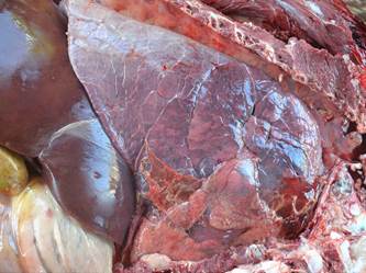
Morbidity and mortality rates varied from 0.5-33.33% and from 27.27-100%, respectively. However, in most of the outbreaks the morbidly rates were below 6% (Fig.1). Cattle from Outbreak 3 were treated with antibiotics every time their rectal temperatures were higher than 39?, which may explain the low lethality rate in this outbreak. Main clinical signs, most frequently affecting muscles, necropsy, and histopathological findings are in Tables 3-6. Clinical signs were observed in 41 cattle; the remaining affected cattle were either found dead (n=10) or no information were registered in their necropsy protocol (n=8). In 20 cases there were no gross description but the histopathological changes were characteristic of blackleg (Table 6) allowing for the diagnosis. Observed gross lesions in striate muscle include locally extensive hemorrhage and edema, often with crepitation of the affected area caused by gas bubbles produced by the organism multiplication. The covering hide was taut and there was a large area of hemorrhage in the subcutaneous tissue, overlying fascia and subcutaneous tissue. Necrotic muscle fibers appeared dark red to red-black (Fig.2). The lesions were wet and exudative (early lesions) or dry (later lesions). A characteristic odor of rancid butter from butyric acid was reported in some in stances. Affected muscle floated in the formalin solution (Fig.3). In case 58 cardiac involvement (Fig.4) and fibrinous pleurits (Fig.5) were observed. Histologically, locally extensive areas of muscle fibers undergoing coagulation necrosis and fragmentation, and interstitial edema and hemorrhage were seen (Fig.6).

Most frequently affected muscles in cases of blackleg diagnosed in cattle from the state of Mato Grosso do Sul from 1994-2014

Gross findings in bovine blackleg. There is a dark hemorrhagic zone within the affected muscle. Necrotic muscle is dark red to red-black. A portion of the lesion to the left is dark red, wet and exudative (early lesions) or dry, red-black with muscle bundles separated by gas bubbles (later lesions).

Gross findings in bovine blackleg. A fragment of an affected muscle floats in 10% formalin solution.


Gross findings in bovine blackleg. Fibrinous pleuritis adhere lung lobes to one another and to pericardial sac.

Histopathological findings in bovine blackleg. Coagulative necrosis of myofibers, hemorrhage and distended bundles of myofibers by gas bubbles. There is mild inflammatory infiltrate of neutrophils. HE, obj.40x.
The laboratory tests used to identify the clostridia causing the myonecrosis and their results are in Table 7. In 12 out of the 17 submitted materials Clostridium Chauvoei was identified as the causative agent.
In Table 8, the economic impact estimated for the outbreaks of the disease is presented. The estimated losses were 0.75 to 27.50% in those farms which did not use vaccination; there was 0.50 to 33.33% loss in farms without information about vaccination, and 0.72 to 6.00% loss in farms where the herd was vaccinated.
The vaccine costs and estimated losses was the following: 15.96 to 0.44% for farms which did not vaccinate, from 36.14 to 0.54% in farms where information on vaccination was not supplied, and from 16.52 to 2.13% in those farms where the herd was vaccinated.
Discussion
Although blackleg is considered an important cause of economic losses in several countries (Useh et al. 2003Useh N.M., Nok A.J. & Esievo K.A.N. 2003. Pathogenesis and pathology of blackleg in ruminants: the role of toxins and neuraminidase, a short review. Vet. Q. 25:155-159., Kriek & Odendaal 2004Kriek N.P.J. & Odendaal M.W. 2004. Clostridium chauvoei infections, p.1856-1862. In: Coetzer R. & Tustin R.C. (Eds), Infectious Diseases of Livestock. Vol.3. 2nd ed. Oxford University Press, Cape Town., Useh et al. 2006bUseh N.M., Ibrahim N.D.G., Nok A.J. & Esievo K.A.N. 2006b. Relationship between outbreaks of blackleg of cattle and annual rainfall in Zaria, Nigeria. Vet. Rec. 158:100-101.) including Brazil (Riet-Correa 2007Riet-Correa F. 2007. Doenças Bacterianas, p.199-443. In: Riet-Correa F., Schild A.L., Lemos R.A.A. & Borges J.R.J. (Eds), Doenças de Ruminantes e Equídeos. Vol. 1. 3ª ed. Pallotti, Santa Maria, RS., Lobato et al. 2013Lobato F.C.F., Salvarani F.M., Gonçalvez L.A., Pires P.S., Silva R.O.S., Alves G.G., Neves M., Oliveira Júnior C.A. & Pereira P.L.L. 2013. Clostridioses dos animais de produção. Vet. Zootec. 20:29-48.), there are few documented surveys on the disease approaching historical series. Cases of blackleg occurred in every year of the time frame of the current study in different regions of the state demonstrating that the pasture contamination by Clostridium chauvoei is disseminated and that the disease is a frequent cause of death in cattle from Mato Grosso do Sul. Considering that C. chauvoei is a soil organism (Kriek & Odendaal 2004Kriek N.P.J. & Odendaal M.W. 2004. Clostridium chauvoei infections, p.1856-1862. In: Coetzer R. & Tustin R.C. (Eds), Infectious Diseases of Livestock. Vol.3. 2nd ed. Oxford University Press, Cape Town., Radostits et al. 2007Radostits O.M., Gay C.C., Hinchcliff K.W. & Constable P.D. 2007. Veterinary medicine: a textbook of the diseases of cattle, horses, sheep, pigs, and goats. 10th ed. W.B. Saunders, Philadelphia. 2156p.) and is widely distributed in this State, it is probable that the low prevalence of cases of blackleg during the period of this study is due to vaccination, a common practice in most farms (Araújo et al. 2010Araújo R.F., Curci V.C.L.M., Nobrega F.L.C., Ferreira R.M.M. & Dutra I.S. 2010. Vaccination protocol and bacterial strain affect the serological response of beef calves against blackleg. Pesq. Vet. Bras. 30(7):554-558.). This is also true in other countries (Uzal 2012Uzal F.A. 2012. Evidence-based medicine concerning efficacy of vaccination against Clostridium chauvoei infection in cattle. Vet. Clin. N. Am., Large Anim. Pract. 28:71-77.). Data from the Syndicate of National Industries of Products for Animal Health, estimates that 150 million of vaccine doses against blackleg are sold annually in Brazil (Araújo et al. 2010Araújo R.F., Curci V.C.L.M., Nobrega F.L.C., Ferreira R.M.M. & Dutra I.S. 2010. Vaccination protocol and bacterial strain affect the serological response of beef calves against blackleg. Pesq. Vet. Bras. 30(7):554-558.).
In the current study blackleg was responsible for 59 deaths out of a total of 5,375 necropsied cattle, thus accounting for 1.1% of the deaths in cattle in studied the period (1994-2014). In a similar study carried out in the South region of Rio Grande do Sul, from 1978-2007 (Fiss et al. 2008Fiss L., Grecco F.B., Ladeira S.R.L., Ferreira J.L.M., Soares M.P., Schramm R., Riet-Correa F. & Schild A.L. 2008. Clostridioses em ruminantes na área de influência do Laboratório Regional de Diagnóstico de 1978-2007. Boletim 28 do Laboratório Regional de Diagnóstico, UFPel, p.31-48.), out of a total of 5,133 cattle examined, there were 29 cases of blackleg, i.e., 0.56% of all the diagnosis in cattle. This made blackleg the most frequent clostridial disease of that part of the country. In another similar study carried out in the Central region of Rio Grande do Sul, from 1964-2008 (Lucena et al. 2010Lucena R.B., Pierezan F., Kommers G.D., Irigoyen L.F., Fighera R.A. & Barros C.S.L. 2010. Doenças de bovinos no Sul do Brasil: 6.706 casos. Pesq. Vet. Bras. 30:428-434.) blackleg was also the most prevalent clostridial disease of cattle, being responsible for 0.63% over the total (6,706) of examined cattle.
With the exception of January, cases of blackleg in cattle in the current study occurred in every month of the year. There was a larger concentration of cases between June-October. Those are similar data are also reported in other regions of Brazil (Fiss et al. 2008Fiss L., Grecco F.B., Ladeira S.R.L., Ferreira J.L.M., Soares M.P., Schramm R., Riet-Correa F. & Schild A.L. 2008. Clostridioses em ruminantes na área de influência do Laboratório Regional de Diagnóstico de 1978-2007. Boletim 28 do Laboratório Regional de Diagnóstico, UFPel, p.31-48.).
Reports from other countries (Kriek & Odendaal 2004Kriek N.P.J. & Odendaal M.W. 2004. Clostridium chauvoei infections, p.1856-1862. In: Coetzer R. & Tustin R.C. (Eds), Infectious Diseases of Livestock. Vol.3. 2nd ed. Oxford University Press, Cape Town., Radostits et al. 2007Radostits O.M., Gay C.C., Hinchcliff K.W. & Constable P.D. 2007. Veterinary medicine: a textbook of the diseases of cattle, horses, sheep, pigs, and goats. 10th ed. W.B. Saunders, Philadelphia. 2156p.) mention seasonality for blackleg occurrence, with most of the cases occurring in the warmer months. These authors admit that variations could occur in the seasonality with most cases occurring during Spring and Fall. Several factors are used to explain the seasonality although none of them is proved.
Most of the cases of blackleg of the current study occurred in cattle from 7-12 months of age, although few younger and older stock were also affected which is similar to what is world widely observed (Kriek & Odendaal 2004Kriek N.P.J. & Odendaal M.W. 2004. Clostridium chauvoei infections, p.1856-1862. In: Coetzer R. & Tustin R.C. (Eds), Infectious Diseases of Livestock. Vol.3. 2nd ed. Oxford University Press, Cape Town., Assis et al. 2005Assis R.A., Facury Filho E.J., Lobato F.C.F., Carvalho A.U., Ferreira P.M. & Carvalho A.V.A. 2005. Surto de carbúnculo sintomático em bezerros. Ciência Rural 35:945-947., Riet-Correa 2007Riet-Correa F. 2007. Doenças Bacterianas, p.199-443. In: Riet-Correa F., Schild A.L., Lemos R.A.A. & Borges J.R.J. (Eds), Doenças de Ruminantes e Equídeos. Vol. 1. 3ª ed. Pallotti, Santa Maria, RS., Groseth et al. 2011Groseth P.K., Ersdal C., Bjelland A.M. & Stokstad M. 2011. Large outbreak of blackleg in housed cattle. Vet. Rec. 169:339-341.). The occurrence of fewer cases in calves younger than 6 month-old is explained by the protection conferred by the colostrum; consequently the occurrence of cases from birth to 6-months-old are attributed to failure in the transfer of passive immunity. Although there are no data explaining the apparent resistance of cattle older than 24-month-old to blackleg, it is probably due to acquired immunity to successive subclinical infection with C. chauvoei. If this is true, then adult cattle may become affected when raised in C. chauvoei-free pastures and transfer when adults into contaminated pasture.
In the current study, in most the outbreak files reviewed, there were no detailed information on vaccination history. In 12 outbreaks calves have not been vaccinated, in 8 outbreaks vaccination has been carried out inadequately and in only two outbreaks vaccination has been done according to the specifications of the manufactures, i.e., the first dose in 4-month-old calves and a boost 30 days after. In three outbreaks calves have been vaccinated twice, when 4-month-old and then when 8-month-old, which can be considered an adequate practice of common use in many farms (Araújo et al. 2010Araújo R.F., Curci V.C.L.M., Nobrega F.L.C., Ferreira R.M.M. & Dutra I.S. 2010. Vaccination protocol and bacterial strain affect the serological response of beef calves against blackleg. Pesq. Vet. Bras. 30(7):554-558.).
One of the difficulties to evaluate whether the protocols were in conformity with the technical recommendations is that most of the farms that do not possess a definite sanitary agenda, in which cattle are set apart in lots according to their ages. It is then possible that the age at which the calves are vaccinated in many farms is markedly variable. For instance, when the first dose is applied at the same time for all the calves in a given farm, calves under and over 4-month-old can be vaccinated. This might be a risky procedure since older calves become susceptible due to the decline of the colostral protection and calves younger than 4-month-old may have lesser protection because maternal-derived antibody will interfere with the ability of young animals to respond to the majority current vaccines (Troxel et al. 1997Troxel R., Burke G.L., Wallace W.T., Keaton L.W., McPeake S.R., Smith D. & Nicholson I. 1997. Clostridial vaccination efficacy on stimulating and maintaining an immune response in beef cows and calves. J. Anim. Sci. 75:19-25., Radostits et al. 2007Radostits O.M., Gay C.C., Hinchcliff K.W. & Constable P.D. 2007. Veterinary medicine: a textbook of the diseases of cattle, horses, sheep, pigs, and goats. 10th ed. W.B. Saunders, Philadelphia. 2156p., Day 2012Day M.J. 2012. Vaccination, p.192-220. In: Idib (Ed.), Veterinary Immunology: principles and practice. Manson Publishing, London.), although this negative effect is no confirmed by some studies (Schipper et al. 1978Schipper I.A., Kelling C.L., Mayer J. & Pfeiffer N.W. 1978. Effects of passive immunity on immune response in calves vaccinated against Clostridium chauvoei infection (blackleg). Vet. Med. Small Anim. Clin. 73:1564-1566.). Furthermore, failure due to differences between vaccinal and field strains of C. chauvoel are reported from Australia (Reed & Reynolds 1977Reed G.A. & Reynolds L. 1977. Failure of Clostridium chauvoei vaccines to protect against blackleg. Aust. Vet. J. 53:393.) and Brazil (Santos 2003Santos B.A. 2003. Avaliação da eficácia em cobaias de imunógenos contra carbúnculo sintomático em uso no Brasil. Dissertação de Mestrado em Medicina Veterinária Preventiva, Faculdade de Ciências Agrárias e Veterinárias, Universidade Estadual Paulista, Jaboticabal, SP. 35p.).
Although vaccination against blackleg is a worldwide disseminated practice there are few reports attesting vaccine efficacy (Uzal 2012Uzal F.A. 2012. Evidence-based medicine concerning efficacy of vaccination against Clostridium chauvoei infection in cattle. Vet. Clin. N. Am., Large Anim. Pract. 28:71-77.). In the current study, the efficacy of vaccination was observed in one of the outbreaks were cattle vaccinated and unvaccinated were kept in the same conditions and only the unvaccinated cattle develop blackleg.
There is wide variation in the lower and upper values for morbidity (0.50 to 33.33%) and lethality (27.27 to 100%) rates in blackleg outbreaks in cattle. Ample variations in these rates have already been reported as 0.66 to 57.14% for morbidity and 85.71 to 100% for lethality (Uzal et al. 2003aUzal F.A., Paramidani M., Assis R., Morris W. & Miyakwa M.F. 2003a. Outbreak of clostridial myocarditis in calves. Vet. Rec. 152:134-136., Assis et al. 2005Assis R.A., Facury Filho E.J., Lobato F.C.F., Carvalho A.U., Ferreira P.M. & Carvalho A.V.A. 2005. Surto de carbúnculo sintomático em bezerros. Ciência Rural 35:945-947., Harwood et al. 2007Harwood D.G., Higgins R.J. & Aggett D.J. 2007. Outbreak of intestinal and lingual Clostridium chauvoei infection in two-year-old Friesian heifers. Vet. Rec. 161:307-308., Groseth et al. 2011Groseth P.K., Ersdal C., Bjelland A.M. & Stokstad M. 2011. Large outbreak of blackleg in housed cattle. Vet. Rec. 169:339-341., Casagrande et al. 2015Casagrande R.A., Pires P.S., Silva R.O.S., Sonne L., Borges J.B.S., Neves M.S., Rolim V.M., Souza S.O., Driemeier D. & Lobato F.C.F. 2015. Histopathological, immunohistochemical and biomolecular diagnosis of myocarditis due to Clostridium chavoei in a bovine. Ciência Rural 45:1472-1475.). In the current study in 22 of 31 outbreaks, morbidly rates were lower than 6% and the higher rates occurred in small cattle herds where only one case would reflect a large percentage. As for the lethality it was less than 100% in 4 of 31 outbreaks. Cautions must be exerted in interpreting these data, because in some instances the outbreak was in progress when observed, and epidemiological data reflect only the time of necropsy since in some outbreaks there was no follow up. It is important to make this disclaimer since blackleg lethality rates are virtually 100% and even treated cattle have poor prognosis (Kriek & Odendaal 2004Kriek N.P.J. & Odendaal M.W. 2004. Clostridium chauvoei infections, p.1856-1862. In: Coetzer R. & Tustin R.C. (Eds), Infectious Diseases of Livestock. Vol.3. 2nd ed. Oxford University Press, Cape Town., Riet-Correa 2007Riet-Correa F. 2007. Doenças Bacterianas, p.199-443. In: Riet-Correa F., Schild A.L., Lemos R.A.A. & Borges J.R.J. (Eds), Doenças de Ruminantes e Equídeos. Vol. 1. 3ª ed. Pallotti, Santa Maria, RS.).
Three cases reported here affected the heart (visceral form) and the remaining affected other striated muscles (classic form). In fact, the classic form usually predominates over the visceral for, the latter being sporadic (Glastonbury et al. 1988Glastonbury J.R.W., Searson J.E., Links I.J. & Tucket L.M. 1988. Clostridial myocarditis in lambs. Aust. Vet. J. 65:208-209., Uzal et al. 2003aUzal F.A., Paramidani M., Assis R., Morris W. & Miyakwa M.F. 2003a. Outbreak of clostridial myocarditis in calves. Vet. Rec. 152:134-136.). Clinical signs, necropsy findings and histopathology observed here are typically the same reported in the literature (Uzal et al. 2003aUzal F.A., Paramidani M., Assis R., Morris W. & Miyakwa M.F. 2003a. Outbreak of clostridial myocarditis in calves. Vet. Rec. 152:134-136., Gregory et al. 2006Gregory L., Della Libera A.M.M., Birgel Júnior E.H., Pogliani F.C., Birgel D.B., Benesi F.J., Miyashiro S. & Baldassi L. 2006. Carbúnculo sintomático: ocorrência, evolução clínica e acompanhamento da recuperação de bovino acometido de “manqueira”. Arqs Inst. Biológico, São Paulo, 73:243-246., Harwood et al. 2007Harwood D.G., Higgins R.J. & Aggett D.J. 2007. Outbreak of intestinal and lingual Clostridium chauvoei infection in two-year-old Friesian heifers. Vet. Rec. 161:307-308., Groseth et al. 2011Groseth P.K., Ersdal C., Bjelland A.M. & Stokstad M. 2011. Large outbreak of blackleg in housed cattle. Vet. Rec. 169:339-341., Casagrande et al. 2015Casagrande R.A., Pires P.S., Silva R.O.S., Sonne L., Borges J.B.S., Neves M.S., Rolim V.M., Souza S.O., Driemeier D. & Lobato F.C.F. 2015. Histopathological, immunohistochemical and biomolecular diagnosis of myocarditis due to Clostridium chavoei in a bovine. Ciência Rural 45:1472-1475., Barros 2016Barros C.S.L. 2016. Sistema muscular, p.663-702. In: Santos R.L. & Alessi A.C. (Eds), Patologia Veterinária. 2ª ed. Roca, São Paulo., Cooper & Valentine 2016Cooper B.J. & Valentine B.A. 2016. Muscle and tendon, p.164-249.In: Maxie M.G. (Ed.), Jubb, Kennedy, and Palmer’s Pathology of Domestic Animals. Vol.1. 6th ed. Elsevier, St Louis.). In the current study the group of muscle most frequently affected was those of the pelvic girdle.
Our results demonstrate that “blackleg diagnosis criteria” used in the current study are reliable for diagnosis of blackleg in cattle. These criteria are used for working diagnosis at several diagnostic laboratories from Brazil (Riet-Correa 2007Riet-Correa F. 2007. Doenças Bacterianas, p.199-443. In: Riet-Correa F., Schild A.L., Lemos R.A.A. & Borges J.R.J. (Eds), Doenças de Ruminantes e Equídeos. Vol. 1. 3ª ed. Pallotti, Santa Maria, RS.). and elsewhere (Kriek & Odendaal 2004Kriek N.P.J. & Odendaal M.W. 2004. Clostridium chauvoei infections, p.1856-1862. In: Coetzer R. & Tustin R.C. (Eds), Infectious Diseases of Livestock. Vol.3. 2nd ed. Oxford University Press, Cape Town., Useh et al. 2006aUseh N.M., Nok A.J. & Esievo K.A. 2006a. Blackleg in ruminants. Perspect. Agric. Vet. Sci. Nutr. Nat. Res. 1:1-8.), due to difficulties in isolating C. chauvoei from samples of clinical cases (Uzal et al. 2003bUzal F.A., Hugenholtz P., Blackall L.L., Petray S., Moss S., Assis R.A., Fernandez Miyakawa M. & Carloni G. 2003b. PCR detection of Clostridium chauvoei in pure cultures and in formalin-fixed, paraffin-embedded tissues. Vet. Microbiol. 91: 239-248.) and due the fact that many times the samples are send to the laboratory fixed in formalin. The use of these criteria proved efficient also in the current study, as 12 of 17 cases diagnosed based “blackleg diagnosis criteria” were confirmed by bacterial culture, PCR or both (Table 7).
The microbiological method for isolation of C. Chauvoei was employed successful in five cases in which the samples were preserved in anaerobiosis environment. In spite of difficulties in the microbiological isolation of the causative agent, this procedure should be encouraged in cases of blackleg in cattle, because cases of vaccination failures due to vaccinal C. chauvoei can occur as already mentioned. Thus, keeping a bank of field strains consist in a measure of security for evaluation of the efficacy of vaccines.
Three samples of cattle fulfilling all the epidemiological and clinicopathological criteria of blackleg were negative for C. Chauvoei in bacterial culture. Two of these samples - from cattle involved in the same outbreak - were stored for 12 months and yield C. septicum in pure culture, which were subsequently confirmed by PCR of the isolates. However all three samples were positive for C. chauvoei DNA by the PCR done in FFPE muscle samples. It is inferred that the storage of material for long periods might favor post mortem proliferation of organisms.
The PCR technique in FFPE was efficient in the identification of C. chauvoei. It detected exclusively C. chauvoei in 8 of 12 cases examined by this technique. Similar results were described by others (Kuhnert et al. 1997Kuhnert P., Krampe M., Capaul S.E., Frey J. & Nicolet J. 1997. Identification of Clostridium chauvoei in cultures and clinical material from blackleg using PCR. Vet. Microbiol. 51:291-298., Uzal et al. 2003bUzal F.A., Hugenholtz P., Blackall L.L., Petray S., Moss S., Assis R.A., Fernandez Miyakawa M. & Carloni G. 2003b. PCR detection of Clostridium chauvoei in pure cultures and in formalin-fixed, paraffin-embedded tissues. Vet. Microbiol. 91: 239-248.). The utilization of PCR in formalin fixed and in FFPE samples is a promising option when the submission to the diagnostic laboratory for bacterial culture cannot be made with the necessary promptness. It is also worth mention that the isolation of C. chauvoei from samples without lesion should not be considered as diagnostic for blackleg, since C. chauvoei can be isolated from healthy cattle (Useh et al. 2006aUseh N.M., Nok A.J. & Esievo K.A. 2006a. Blackleg in ruminants. Perspect. Agric. Vet. Sci. Nutr. Nat. Res. 1:1-8.). Several clostridial organisms including C. perfringens, C. septicum, C. sordellii are saprophytes of the intestine of cattle and can proliferate and disseminate in the body during the terminal stages or after the animal death from an unrelated disease. Thus the isolation of pathogenic clostridia from a necropsy case is of difficult interpretation and cannot be diagnostic unless the lesions compatible with the suspect disease are present or the toxin is demonstrated by tests in mice (Hjerpe 1990Hjerpe C.A. 1990. Bovine vaccines and herd vaccination programs. Vet. Clin. N. Am., Food Anim. Pract. 6:171-214.).
The difficult in isolation of C. chauvoei reinforces the importance of necropsy and histopathology for the diagnosis of blackleg. Several factors may interfere with the bacteriological diagnosis, such as time elapsed from the sampling of material and the performance of the analysis and also on conservation and non-contamination of the collected sample (Radostits et al. 2007Radostits O.M., Gay C.C., Hinchcliff K.W. & Constable P.D. 2007. Veterinary medicine: a textbook of the diseases of cattle, horses, sheep, pigs, and goats. 10th ed. W.B. Saunders, Philadelphia. 2156p., Riet-Correa 2007Riet-Correa F. 2007. Doenças Bacterianas, p.199-443. In: Riet-Correa F., Schild A.L., Lemos R.A.A. & Borges J.R.J. (Eds), Doenças de Ruminantes e Equídeos. Vol. 1. 3ª ed. Pallotti, Santa Maria, RS.).
Clostridium septicum is a postmortem invasor promoting putrefaction of the carcass, and may be present at the time of tissue sampling not necessarily being involved in the pathogenesis of the disease investigated (Kuhnert et al 1997Kuhnert P., Krampe M., Capaul S.E., Frey J. & Nicolet J. 1997. Identification of Clostridium chauvoei in cultures and clinical material from blackleg using PCR. Vet. Microbiol. 51:291-298., Lobato et al. 2007Lobato F.C.F., Salvarani F.M. & Assis R.A. 2007. Clostridioses dos pequenos ruminantes. Revta Port. Ciênc. Vet. 102:23-34.). Furthermore C. septicum inhibits the growth of do C. chauvoei in solid culture medium. Co-infections with C. chauvoei and C. septicum are not uncommon; however, the importance of C. septicum as etiologic agent in bovine blackleg is controversial (Radostits et al. 2007Radostits O.M., Gay C.C., Hinchcliff K.W. & Constable P.D. 2007. Veterinary medicine: a textbook of the diseases of cattle, horses, sheep, pigs, and goats. 10th ed. W.B. Saunders, Philadelphia. 2156p.).
In the current study C. chauvoei and C. septicum were not cultured from the same lesion; however in two cases in which only C. septicum was cultured, C. chauvoei was identified by the PCR technique in FFPE. In a study involving 176 cases of clostridial myonecrosis in cattle, C. chauvoei was found alone or associated with C. septicum in 56% of the cases and in 36% of the cases C. novyi or C. novyi and C. septicum were cultured from the lesion (Williams 1977Williams B.M. 1977. Clostridial myositis in cattle: bacteriology and gross pathology. Vet. Rec. 100(5):90-91.). Although some authors consider that these results indicate that for the best protection of a cattle herd one should use polyvalent vaccines containing all these antigens (Radostits et al. 2007Radostits O.M., Gay C.C., Hinchcliff K.W. & Constable P.D. 2007. Veterinary medicine: a textbook of the diseases of cattle, horses, sheep, pigs, and goats. 10th ed. W.B. Saunders, Philadelphia. 2156p.), in Brazil, field observations point to C. chauvoei as the sole cause, since for many years large cattle herds were successfully immunized by monovalent vaccines (Lobato et al. 2004Lobato F.C.F., Assis R.A., Balsamão G.M., Abreu V.L.V., Nascimento R.A.P. & Neves R.D. 2004. Eficácia de vacinas comerciais contra clostridioses frente ao desafio com Clostridium sordellii. Ciência Rural 34:439-442.).
The results of the current study demonstrate that blackleg is a frequent disease in cattle from Mato Grosso do Sul and that it could be responsible for significant economic losses to farmers. The estimated losses, costs and economic impact stemming from the outbreaks of blackleg demonstrate that herds not vaccinated or vaccinated incorrectly elevate the losses and impact in the farmers profit since cattle blackleg has a high lethality rates. There is scarcity of data compromising the more accurate assessment of the economic impact. The cost of these losses may be reduced by the installment of an adequate vaccination program that takes into account efficient vaccines against the C. chauvoei strains prevalent in the region, and abiding to the protocols indicated by the manufacturers.
Conclusions
Bovine blackleg is a frequent disease in Mato Grosso does Sul, occurring all year round. Most affected cattle are 7 to 12-month old calves, although sporadic cases might occur in cattle younger than 6 and older than 14-month old.
The classic form predominates, but a small percentage of visceral blackleg occur.
The number of blackleg cases in properly vaccinated cattle herds was lower than the one in not vaccinated cattle or in those vaccinated incorrectly.
Acknowledgments
The authors are indebted to Prof. Claudio Barros for helping with the editing the manuscript and translating it English. Prof. Barros is a Visiting Professor at the Faculty of Veterinary Medicine and Animal Husbandry and is funded through a fellowship from FUNDECT/CAPES(052014PVMS59/300.032/2015).
References
- Araújo R.F., Curci V.C.L.M., Nobrega F.L.C., Ferreira R.M.M. & Dutra I.S. 2010. Vaccination protocol and bacterial strain affect the serological response of beef calves against blackleg. Pesq. Vet. Bras. 30(7):554-558.
- Assis R.A., Facury Filho E.J., Lobato F.C.F., Carvalho A.U., Ferreira P.M. & Carvalho A.V.A. 2005. Surto de carbúnculo sintomático em bezerros. Ciência Rural 35:945-947.
- Barros C.S.L. 2016. Sistema muscular, p.663-702. In: Santos R.L. & Alessi A.C. (Eds), Patologia Veterinária. 2ª ed. Roca, São Paulo.
- Burke M.P. & Opeskin K. 1999. Non traumatic clostridial myonecrosis. Am. J. Forensic Med. Pathol. 20:158-162.
- Casagrande R.A., Pires P.S., Silva R.O.S., Sonne L., Borges J.B.S., Neves M.S., Rolim V.M., Souza S.O., Driemeier D. & Lobato F.C.F. 2015. Histopathological, immunohistochemical and biomolecular diagnosis of myocarditis due to Clostridium chavoei in a bovine. Ciência Rural 45:1472-1475.
- Cooper B.J. & Valentine B.A. 2016. Muscle and tendon, p.164-249.In: Maxie M.G. (Ed.), Jubb, Kennedy, and Palmer’s Pathology of Domestic Animals. Vol.1. 6th ed. Elsevier, St Louis.
- Day M.J. 2012. Vaccination, p.192-220. In: Idib (Ed.), Veterinary Immunology: principles and practice. Manson Publishing, London.
- Fiss L., Grecco F.B., Ladeira S.R.L., Ferreira J.L.M., Soares M.P., Schramm R., Riet-Correa F. & Schild A.L. 2008. Clostridioses em ruminantes na área de influência do Laboratório Regional de Diagnóstico de 1978-2007. Boletim 28 do Laboratório Regional de Diagnóstico, UFPel, p.31-48.
- Glastonbury J.R.W., Searson J.E., Links I.J. & Tucket L.M. 1988. Clostridial myocarditis in lambs. Aust. Vet. J. 65:208-209.
- Gregory L., Della Libera A.M.M., Birgel Júnior E.H., Pogliani F.C., Birgel D.B., Benesi F.J., Miyashiro S. & Baldassi L. 2006. Carbúnculo sintomático: ocorrência, evolução clínica e acompanhamento da recuperação de bovino acometido de “manqueira”. Arqs Inst. Biológico, São Paulo, 73:243-246.
- Groseth P.K., Ersdal C., Bjelland A.M. & Stokstad M. 2011. Large outbreak of blackleg in housed cattle. Vet. Rec. 169:339-341.
- Harwood D.G., Higgins R.J. & Aggett D.J. 2007. Outbreak of intestinal and lingual Clostridium chauvoei infection in two-year-old Friesian heifers. Vet. Rec. 161:307-308.
- Hatheway C.L. 1990. Toxigenic Clostridia. Clin. Microbiol. Rev. 3:66-98.
- Hjerpe C.A. 1990. Bovine vaccines and herd vaccination programs. Vet. Clin. N. Am., Food Anim. Pract. 6:171-214.
- Kojima A., Uchida I., Sekizaki T., Sasaki Y., Ogikubo Y. & Tamura Y. 2001. Rapid detection and identification of Clostridium chauvoei by PCR based on flagellin gene sequence. Vet. Microbiol. 78:363-371.
- Kriek N.P.J. & Odendaal M.W. 2004. Clostridium chauvoei infections, p.1856-1862. In: Coetzer R. & Tustin R.C. (Eds), Infectious Diseases of Livestock. Vol.3. 2nd ed. Oxford University Press, Cape Town.
- Kuhnert P., Krampe M., Capaul S.E., Frey J. & Nicolet J. 1997. Identification of Clostridium chauvoei in cultures and clinical material from blackleg using PCR. Vet. Microbiol. 51:291-298.
- Lobato F.C.F., Assis R.A., Balsamão G.M., Abreu V.L.V., Nascimento R.A.P. & Neves R.D. 2004. Eficácia de vacinas comerciais contra clostridioses frente ao desafio com Clostridium sordellii Ciência Rural 34:439-442.
- Lobato F.C.F., Salvarani F.M. & Assis R.A. 2007. Clostridioses dos pequenos ruminantes. Revta Port. Ciênc. Vet. 102:23-34.
- Lobato F.C.F., Salvarani F.M., Gonçalvez L.A., Pires P.S., Silva R.O.S., Alves G.G., Neves M., Oliveira Júnior C.A. & Pereira P.L.L. 2013. Clostridioses dos animais de produção. Vet. Zootec. 20:29-48.
- Lucena R.B., Pierezan F., Kommers G.D., Irigoyen L.F., Fighera R.A. & Barros C.S.L. 2010. Doenças de bovinos no Sul do Brasil: 6.706 casos. Pesq. Vet. Bras. 30:428-434.
- Maxie M.G. & Miller M.A. 2016. Introduction to the diagnostic process, p.1-15. In: Maxie M.G. (Ed.), Jubb, Kennedy, and Palmer’s Pathology of Domestic Animals. Vol. 1. 6th ed. Elsevier, St Louis .
- Radostits O.M., Gay C.C., Hinchcliff K.W. & Constable P.D. 2007. Veterinary medicine: a textbook of the diseases of cattle, horses, sheep, pigs, and goats. 10th ed. W.B. Saunders, Philadelphia. 2156p.
- Reed G.A. & Reynolds L. 1977. Failure of Clostridium chauvoei vaccines to protect against blackleg. Aust. Vet. J. 53:393.
- Ribeiro M.G., Silva R.O.S., Pires P.S., Martinho A.P.V., Lucas T.M., Teixeira A.I.P., Paes A.C., Barros C.B. & Lobato F.C.F. 2012. Myonecrosis by Clostridium septicum in a dog, diagnosed by a new multiplex-PCR. Anaerobe 18:504-507.
- Riet-Correa F. 2007. Doenças Bacterianas, p.199-443. In: Riet-Correa F., Schild A.L., Lemos R.A.A. & Borges J.R.J. (Eds), Doenças de Ruminantes e Equídeos. Vol. 1. 3ª ed. Pallotti, Santa Maria, RS.
- Santos B.A. 2003. Avaliação da eficácia em cobaias de imunógenos contra carbúnculo sintomático em uso no Brasil. Dissertação de Mestrado em Medicina Veterinária Preventiva, Faculdade de Ciências Agrárias e Veterinárias, Universidade Estadual Paulista, Jaboticabal, SP. 35p.
- Schipper I.A., Kelling C.L., Mayer J. & Pfeiffer N.W. 1978. Effects of passive immunity on immune response in calves vaccinated against Clostridium chauvoei infection (blackleg). Vet. Med. Small Anim. Clin. 73:1564-1566.
- Troxel R., Burke G.L., Wallace W.T., Keaton L.W., McPeake S.R., Smith D. & Nicholson I. 1997. Clostridial vaccination efficacy on stimulating and maintaining an immune response in beef cows and calves. J. Anim. Sci. 75:19-25.
- Useh N.M., Nok A.J. & Esievo K.A.N. 2003. Pathogenesis and pathology of blackleg in ruminants: the role of toxins and neuraminidase, a short review. Vet. Q. 25:155-159.
- Useh N.M., Nok A.J. & Esievo K.A. 2006a. Blackleg in ruminants. Perspect. Agric. Vet. Sci. Nutr. Nat. Res. 1:1-8.
- Useh N.M., Ibrahim N.D.G., Nok A.J. & Esievo K.A.N. 2006b. Relationship between outbreaks of blackleg of cattle and annual rainfall in Zaria, Nigeria. Vet. Rec. 158:100-101.
- Uzal F.A., Paramidani M., Assis R., Morris W. & Miyakwa M.F. 2003a. Outbreak of clostridial myocarditis in calves. Vet. Rec. 152:134-136.
- Uzal F.A., Hugenholtz P., Blackall L.L., Petray S., Moss S., Assis R.A., Fernandez Miyakawa M. & Carloni G. 2003b. PCR detection of Clostridium chauvoei in pure cultures and in formalin-fixed, paraffin-embedded tissues. Vet. Microbiol. 91: 239-248.
- Uzal F.A. 2012. Evidence-based medicine concerning efficacy of vaccination against Clostridium chauvoei infection in cattle. Vet. Clin. N. Am., Large Anim. Pract. 28:71-77.
- Valentine B.A. & McGavin M.D. 2012. Skeletal muscle, p.871-919. In: Zachary F.J. & McGavin M.D. (Eds), Pathologic Basis of Veterinary Disease. 5th ed. Elsevier, St Louis .
- Williams B.M. 1977. Clostridial myositis in cattle: bacteriology and gross pathology. Vet. Rec. 100(5):90-91.
Publication Dates
-
Publication in this collection
Jan 2018
History
-
Received
16 Aug 2016 -
Accepted
17 Mar 2017