Abstracts
To establish reference values and to assess the influence of age on the leukograms of healthy Holstein calves, blood samples were obtained from 300 animals. These samples were distributed equally (n=20) among 15 experimental groups according to age: birth to 8 hours, 9 to 16 hours, 17 to 24 hours, 2 days, 3 days, 4 days, 5 days, 6 to 7 days, 8 to 9 days, 10 to 11 days, 12 to 13 days, 14 to 15 days, 16 to 20 days, 21 to 25 days and 26 to 30 days of age. The maximum numbers of leukocytes (9,305.0/mL), segmented neutrophils (6,551.2/mL) and total neutrophils (6,678.3/mL) were noted within the first 8 hours of life, while band neutrophils peaked in number (133.3/mL) between 9 and 16 hours after birth. Meanwhile, the maximum total lymphocyte (4,992.1/µL) and typical lymphocyte (4,686.1/µL) counts occurred between 21 and 25 days, whereas atypical lymphocytes (388.5/µL) reached their maximum number between 26 and 30 days, demonstrating an inversion of the neutrophil:lymphocyte ratio over time. Thus, the influence of age on the leukocyte count of the evaluated calves was verified. The release of endogenous corticosteroids during labor or at birth may contribute to this variation in leukograms with age.
Calves; leukogram; Holstein cattle
Com a finalidade de estabelecer os valores de referência e de avaliar a influência do fator etário sobre o leucograma de bezerras sadias, da raça Holandesa, utilizaram-se amostras de sangue de 300 animais, distribuídos com igual número (n=20) por 15 grupos experimentais, de acordo com a idade: do nascimento até 8 horas de vida; de 9 até 16 horas; de 17 até 24 horas; 2 dias de idade; 3 dias; 4 dias; 5 dias; 6 a 7 dias; 8 a 9 dias; 10 a 11 dias; 12 a 13 dias; 14 a 15 dias; de 16 a 20 dias; de 2l a 25 dias; e de 26 a 30 dias de vida. Puderam-se observar valores máximos nas primeiras oito horas de vida para leucócitos (9305,0/µL), neutrófilos segmentados (6551,2/µL) e neutrófilos totais (6678,3/µL); entre 9 a 16 horas pós-nascimento para os neutrófilos bastonetes (133,3/µL). Para os linfócitos totais (4992,1/µL) e os linfócitos típicos (4686,1/µL) os valores máximos ocorreram entre 21 e 25 dias, e para os linfócitos atípicos (388,5/µL), entre 26 e 30 dias, demonstrando-se uma inversão na relação neutrófilo: linfócito, com o decorrer dos dias de vida. Constatou-se a da influência do fator etário no leucograma dos bezerros avaliados, no entanto acredita-se que a liberação de corticoides endógenos no momento do parto/nascimento, contribuíram para esta variação.
Bezerras; leucograma; gado holandês
ANIMAL MORPHOPHYSIOLOGY
Leukograms of healthy Holstein calves within the first month of life
Leucograma de bezerros Holstein sadios no primeiro mês de vida
Fernando J. BenesiI, * * Corresponding author: febencli@usp.br ; Cynthia M.C. TeixeiraII; Marta L.R. LealII; Julio A.N. LisboaII; Regina M.S. MirandolaI; Carolina L. ShecairaII; Viviani GomesI
IDepartamento de Clínica Médica, Faculdade de Medicina Veterinária e Zootecnia (FMVZ), Universidade de São Paulo (USP), Avenida Prof. Dr. Orlando Marques de Paiva 87, São Paulo, SP 05508000, Brazil
IIPostgraduate Program in Veterinary Medicine, Departamento de Clínica Médica, FMVZ-USP, São Paulo, SP
ABSTRACT
To establish reference values and to assess the influence of age on the leukograms of healthy Holstein calves, blood samples were obtained from 300 animals. These samples were distributed equally (n=20) among 15 experimental groups according to age: birth to 8 hours, 9 to 16 hours, 17 to 24 hours, 2 days, 3 days, 4 days, 5 days, 6 to 7 days, 8 to 9 days, 10 to 11 days, 12 to 13 days, 14 to 15 days, 16 to 20 days, 21 to 25 days and 26 to 30 days of age. The maximum numbers of leukocytes (9,305.0/mL), segmented neutrophils (6,551.2/mL) and total neutrophils (6,678.3/mL) were noted within the first 8 hours of life, while band neutrophils peaked in number (133.3/mL) between 9 and 16 hours after birth. Meanwhile, the maximum total lymphocyte (4,992.1/µL) and typical lymphocyte (4,686.1/µL) counts occurred between 21 and 25 days, whereas atypical lymphocytes (388.5/µL) reached their maximum number between 26 and 30 days, demonstrating an inversion of the neutrophil:lymphocyte ratio over time. Thus, the influence of age on the leukocyte count of the evaluated calves was verified. The release of endogenous corticosteroids during labor or at birth may contribute to this variation in leukograms with age.
Index terms: Calves, leukogram, Holstein cattle.
RESUMO
Com a finalidade de estabelecer os valores de referência e de avaliar a influência do fator etário sobre o leucograma de bezerras sadias, da raça Holandesa, utilizaram-se amostras de sangue de 300 animais, distribuídos com igual número (n=20) por 15 grupos experimentais, de acordo com a idade: do nascimento até 8 horas de vida; de 9 até 16 horas; de 17 até 24 horas; 2 dias de idade; 3 dias; 4 dias; 5 dias; 6 a 7 dias; 8 a 9 dias; 10 a 11 dias; 12 a 13 dias; 14 a 15 dias; de 16 a 20 dias; de 2l a 25 dias; e de 26 a 30 dias de vida. Puderam-se observar valores máximos nas primeiras oito horas de vida para leucócitos (9305,0/µL), neutrófilos segmentados (6551,2/µL) e neutrófilos totais (6678,3/µL); entre 9 a 16 horas pós-nascimento para os neutrófilos bastonetes (133,3/µL). Para os linfócitos totais (4992,1/µL) e os linfócitos típicos (4686,1/µL) os valores máximos ocorreram entre 21 e 25 dias, e para os linfócitos atípicos (388,5/µL), entre 26 e 30 dias, demonstrando-se uma inversão na relação neutrófilo: linfócito, com o decorrer dos dias de vida. Constatou-se a da influência do fator etário no leucograma dos bezerros avaliados, no entanto acredita-se que a liberação de corticoides endógenos no momento do parto/nascimento, contribuíram para esta variação.
Termos de indexação: Bezerras, leucograma, gado holandês.
INTRODUCTION
Adaptation to the extrauterine environment is crucial to the survival of calves during the neonatal period, particularly because their immune systems are still developing (Benesi 1992). The diseases most often affecting calves are diarrhea, umbilical complications, pneumonia and septicemia, which can cause hematological alterations in addition to specific clinical symptoms. Research on these diseases and the corresponding hematological changes is critical, as a reduction in such illnesses may diminish or prevent the large economic losses associated with the death of calves, and especially of females with good milk-producing characteristics and of high genetic merit.
The clinical evaluation of sick neonates, with the goals of diagnosis, determination of prognosis and treatment, often requires the combination of a physical examination and auxiliary laboratory tests. The leukogram is a key tool in patient evaluation, either confirming or elucidating the diagnosis of a disease and thus guiding therapy and prognosis prediction (Benesi 1992, 1993).
The proper interpretation of the results obtained from the combination of a physical examination and laboratory tests requires collection of the maximum amount of information possible during the examination, as well as knowledge of the physiology of the affected organs and systems and the pathogenesis of the disease. An additional crucial requirement is having specific reference ranges that account for the species, breed, age, gender and environmental/climatic factors. Although it is known that precise and justified interpretation of leukograms requires reference ranges, few studies of these ranges have been conducted for bovine neonates (Benesi 1992, Biondo 1996, Fagliari et al. 1998).
Past studies reported that the mean leukocyte numbers in the leukograms of calves assessed immediately after birth did not change significantly, although some specific trends were highlighted. Within the first six hours after birth (a.b.), a small decrease in the total number of leukocytes was noted, while neutrophils were predominant and the absolute number of lymphocytes reached a minimum. A decrease in the absolute numbers of eosinophils and monocytes was also noted, with minimum values achieved 12 hours a.b. After this time-point, an increase in total leukocytes was observed due to a similar increase in the total number of neutrophils, especially segmented neutrophils (Birgel 1972, La Motte & Eberhart 1976), and in the number of lymphocytes and monocytes (Birgel 1972, Tennant et al. 1974, Benesi 1992). A high neutrophil:lymphocyte ratio (Ne:Ly) was also noted at birth, as well as a reduction in or inversion of this ratio within the first week of life due to the numerical predominance of lymphocytes (Adams et al. 1992, Biondo 1996, Costa et. al. 2008).
Immature, young and band neutrophils in the circulating blood of calves during the first week of life have been reported by some authors (Anders 1966, Benesi 1992). The changes observed in the calves' leukograms within the first week of life have been attributed to the stress of labor and birth, as reflected by the elevated levels of circulating endogenous glucocorticoids and their effects on leukocyte behavior (Eberhart & Patt 1971, La Motte & Eberhart 1976, Benesi 1992). Particularly during the neonatal period, variations in leukograms may occur as a result of the type of labor and the intensity of the obstetric maneuvers (Birgel 1972, Benesi 1992), as well as a result of the health of the mother and the neonate. In addition to being influenced by the birthing process and the feeding of the colostrum, the complete blood count (CBC) may vary due to such factors as age, gender, breed, environment and management practices. The existence of these factors justifies the need to establish reference values for each country or location where the animals are reared (Birgel 1972, 1982, Tennant et al. 1974, Biondo 1996). Therefore, the present study sought to evaluate the leukograms of healthy Holstein calves from Grade A and Grade B dairy farms, with careful consideration of the first month of life of these neonates.
MATERIALS AND METHODS
Three hundred healthy Holstein calves of up to 30 days of age, originating from Grade A and Grade B dairy farms located in the State of São Paulo, Brazil, were included in the current study. The calves were divided into 15 experimental groups of 20 animals each, according to age range: birth to 8 hours, 9 to 16 hours, 17 to 24 hours, 2 days, 3 days, 4 days, 5 days, 6 to 7 days, 8 to 9 days, 10 to 11 days, 12 to 13 days, 14 to 15 days, 16 to 20 days, 21 to 25 days, and 26 to 30 days of age.
The calves selected for this experiment had not yet been subjected to the pre-immunization program for Babesia spp. and Anaplasma sp. and were considered clinically healthy following a physical examination (Dirksen et al. 1993). Any animals exhibiting a packed cell volume below 25% or above 40% were excluded from the study, as well as those exhibiting a total leukocyte count outside of the range of 8-12x103/µl or those infected with blood parasites.
Blood samples were obtained by external jugular venipuncture using a vacuum blood-collection system and a silicone tube containing tripotassium EDTA at a ratio of 1.5 mg/mL of blood. Leukocyte counts were then performed in a modified Neubauer chamber and expressed in thousands of cells per microliter of blood, with Thomas fluid as the diluent. The differential leukocyte count was performed on stained blood smears, with analysis of the differentiation of 100 white blood cells, which allowed for the classification of typical and atypical lymphocytes (Birgel 1982, Jain 2000).
A statistical analysis was performed assuming a completely randomized design with a single factor (age). The effect of age and the interaction between two factors was tested by an analysis of variance. When the F-statistics were significant, Tukey's test was used to verify the differences between the means, calculating the minimum significant difference and assuming an error probability of 5%. All means and standard deviations are shown as measures of central tendency and dispersion for the studied variables.
RESULTS
Throughout the study, blood samples were collected from 385 calves, of which 85 samples were excluded due to the presence of hematologic alterations that indicated anemia, hemoconcentration, leukocytosis, leukopenia or the presence of blood parasites.
Of the 22 dairy farms included in the current study, 7 produced Grade A milk and 15 produced Grade B milk. The farms were located in 14 municipalities of the State of São Paulo: Analândia, Araras, Campinas, Descalvado, Itatiba, Monte Mor, Pindamonhangaba, Pirassununga, Santa Rita do Passa Quatro, São Carlos, São José dos Campos, Tambaú, Tapiratiba and Tietê.
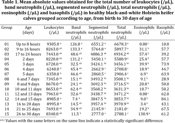
The results obtained in this study are shown in Tables 1 and 2. The total leukocyte counts of the studied animals fluctuated within a small range up to 8 hours a.b. The same fluctuation was noted for the segmented and total neutrophils. The band neutrophils achieved their maximum mean values between 9 and 16 hours of life, followed by a sharp decline, with some instability, that lasted until the 30th day a.b. Meanwhile, the basophils and eosinophils were absent during the first 8 hours a.b., although their levels also fluctuated throughout the period studied, with the eosinophils reaching their highest values at 26 to 30 days of age (Table 1).
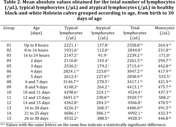
The total and typical lymphocytes counts reached their maximum mean values between 21 and 25 days of age and their minimum values from 9 to 16 hours a.b. In turn, the maximum atypical lymphocyte counts were noted between 26 and 30 days of age, while the minimum values occurred between 17 and 24 hours a.b. (Table 2).
DISCUSSION
The analysis of the mean total leukocyte count showed that its magnitude was greatest in animals between 0 and 8 hours of age and subsequently significantly decreased until its minimum at four days a.b. The values then increased and almost returned to the baseline between 14 and 20 days of age.
A comparison of the mean values observed for all of the studied age groups with the values reported in the non-Brazilian studies for calves of similar ages demonstrated that the counts obtained here were similar in most cases (Kraft 1967, La Motte & Eberhart 1976, Lumsden et al. 1980) or lower (Greatorex 1954, Holman 1956, Kaneko & Mills 1970). Meanwhile, a comparison with results obtained in Brazilian studies conducted on Holstein cattle of up to 1 year of age (Garcia 1989) or up to 3 months of age (Tavora 1997) showed that the data reported here are smaller in magnitude, confirming the need to specifically study bovine neonates.
The changes in the total leukocyte count reflected the behavior of some of the different types of leukocytes. For example, the variation in the mean total neutrophil count was influenced by age and characterized by maximum values between birth and 8 hours of life, followed by a significant decrease beginning at 3 days a.b. and the maintenance of the lower values, with small oscillations, until 30 days of age. The evaluation of the variation in the mean total lymphocyte count also demonstrated the influence of age, although the trend was opposite to that observed for the total number of neutrophils. The minimum mean total lymphocyte count was noted between 9 and 16 hours of life, followed by a gradual and significant increase between 6 and 7 days of age; this increase was maintained, with small fluctuations, until almost 30 days of age, with the maximum value observed between 21 and 25 days a.b.
Considering the simultaneous variation in the total numbers of leukocytes, neutrophils and lymphocytes, it may be hypothesized that the total leukocyte count initially reflected both the increased number of neutrophils and the diminished number of lymphocytes. Therefore, leukocytosis was not observed during the first day of life, which has been described by several authors, although they demonstrated a maximum total leukocyte count due to a similar increase observed in the total neutrophil count (Greatorex 1954, Holman 1956, Kaneko & Mills 1970, Birgel 1972, Tennant et al. 1974, La Motte & Eberhart 1976, Adams et al. 1992, Benesi 1992). The changes in the total neutrophil number particularly reflected the variation observed in the segmented neutrophil count. Thus, the latter type of polymorphonuclear cell was likely responsible for the elevation in the total number of neutrophils, confirming the findings of Birgel (1972), Tennant et al. (1974), Jain (1986), Adams et al. (1992), Benesi (1992) and Biondo (1996). Meanwhile, the failure to detect immature, young and band neutrophils in the circulating blood of the studied calves during the first week of life is consistent with the observation of these cells only by very few authors (Anders 1966, Benesi 1992).
A concomitant assessment of the ratio of total neutrophils:total lymphocytes in the first three days a.b. reveals the predominance of polymorphonuclear cells and shows that this relationship was inverted at four days of age, although this inversion was not sustained. However, the inversion returned after seven days of age, when the numerical predominance of lymphocytes became apparent. Similar observations have been reported by Holman (1956), Kraft (1967), Tennant et al. (1974), Adams et al. (1992) and Benesi (1992).
Changes in the leukograms of calves within the first week of life are thought to be due to the stress of labor or birth. This stress is suggested by the elevation in circulating levels of endogenous glucocorticoids in the blood of these animals and their effects on different blood leukocytes, which are characterized by lymphopenia and eosinopenia; by the absence of eosinophils, which is associated with neutrophilia and monocytosis; or by a normal number of monocytes, in addition to a normal, increased or decreased number of total leukocytes resulting from these changes, depending on whether the normal leukocyte count had a predominance of lymphocytes or neutrophils (Eberhart & Patt 1971, Tennant et al. 1974, La Motte & Eberhart 1976, Benesi 1992). The bovine bone marrow does not possess a large reserve of mature neutrophils, which may explain why neutrophilia is not be sufficiently intense to overcome lymphopenia and the increased total leukocyte count. However, immature neutrophils can rapidly reach the blood in response to cortisol stimulation (Eberhart & Patt 1971, Jain 2000). Although the presence of immature neutrophils suggests inflammation, it may indicate a normal physiological response in neonatal calves to increases in the serum levels of the cortisol associated with labor (Adams et al. 1992, Benesi 1992).
In the post-birth period, additional changes in the endogenous cortisol levels may be influenced by the type of labor and the intensity of the obstetric maneuvers (Birgel 1972, Benesi, 1992). The endogenous cortisol levels may also be affected by the health of the mother and the neonate, which is a determinant of the degree of stress experienced by the neonate.
In general, the changes in the total numbers of leukocytes, neutrophils and lymphocytes observed in the present study are similar to those occurring under the influence of elevated serum endogenous corticosteroid levels. Therefore, these changes may be considered to be a consequence of the stress of birth.
The classification of lymphocytes into typical and atypical, according to their morphological and staining characteristics, was performed in order to verify their numerical variation, given that no reference values have been eslished for dairy-cattle neonates. The typical lymphocytes, which include large and small lymphocytes, exhibited lower numbers between birth and 3 days of age, subsequently evolving into a significant increase from 8 to 9 days of age and reaching a maximum value between 21 and 25 days of age. The typical lymphocyte count was thus mainly responsible for the variation in the mean total lymphocyte count.
The atypical lymphocytes, which encompass monocytoid lymphocytes, plasmacytoid or Turk cells, Gumprecht cells or nuclear shadows and lymphocytes with double nuclei and with pyknotic nuclei, reached a minimum mean total count in the first day of age. The count then increased significantly from 6 to 7 days of life and attained a maximum value between 26 and 30 days a.b.
The monocytes varied in their mean values, achieving a minimum between 17 and 24 hours of life, followed by a moderate increase until a maximum and significantly greater value was measured at five days of age. The monocyte numbers then stabilized, with small fluctuations, until 30 days of age. These observations are in agreement with those of Greatorex (1954) and Tennant et al. (1974) and are discordant with those of Adams et al. (1992). The latter report verified a decrease in monocytes between birth and 24 hours of life, followed by an increase, with values reaching those for adult animals in the third week of life. Most authors reported a small change in the number of monocytes, as observed in the present study up to 30 days of age in calves (Holman 1956, Kaneko & Mills 1970, Benesi 1992, Biondo 1996, Fagliari et al. 1998).
The mean eosinophil and basophil counts observed among the age groups of calves in the present study were always small and fluctuated slightly between birth and 30 days of age. No statistically significant change was noted in the basophil count, consistent with the findings of Birgel (1972), Adams et al. (1992) and Benesi (1992). The small variation in the numbers of eosinophils and basophils has also been reported in previous studies (Tennant et al. 1974, Biondo 1996, Fagliari et al. 1998), while others have identified an increase in the numbers with age ( Holman 1956, Biondo 1996, Fagliari et al. 1998). In this study, the basophil count also increased with age, and a significant increase in eosinophils to the maximum value was noted between 26 and 30 days of age. According to Eberhart & Patt (1971), Tennant et al. (1974) and Adams et al. (1992), eosinophils and basophils are not often observed at birth, possibly due to the increased cortisol concentration in the circulation. However, these two cell types are present in small numbers in the blood of both young calves and adult animals.
CONCLUSION
The maximum mean total leukocyte and neutrophil counts and the concomitant smaller total numbers of lymphocytes and diminished numbers or absence of eosinophils were observed during the first three days of age in many of the studied calves. Thus, despite the influence of age, leukograms observed early in life are consistent with the predominant influence of high levels of endogenous corticosteroids resulting from the stress of birth.
The leukograms of calves in the first week of life should be carefully interpreted, since they are distinct from those of healthy older animals which present a predominance of polymorphonuclear neutrophils mostly comprising segmented cells.
Received on December 2, 2011.
Accepted for publication on January 11, 2012.
- Adams R.R., Garry F.B. & Aldribge B.M. 1992. Haematologic values in newborn beef calves. Am. J. Vet. Res. 53(6):944-50.
- Anders A. 1966. Ein Beitrag zum Blutbild des Kalbes. Dissertation, Fakultät für Veterinärmedizin, Berlin. 100p.
- Benesi F.J. 1992. Hematologia de bezerros recém-nascidos. Influência da asfixia neonatal, do tipo do parto e da ingestão de colostro sobre a crase sanguínea. Tese de Doutorado, Faculdade de Medicina Veterinária e Zootecnia, Universidade de São Paulo, São Paulo, SP. 126p.
- Benesi F.J. 1993. Síndrome asfixia neonatal dos bezerros: importância e avaliação crítica. Arqs Esc. Med. Vet. UFBA, Salvador, 16(1):38-48.
- Biondo A.W. 1996. Hemograma de bovinos sadios da raça Nelore no primeiro mês de vida, criados no estado de São Paulo: influência de fatores etários e sexuais. Dissertação de Mestrado, Universidade Federal de Santa Maria, Santa Maria, RS. 76p.
- Birgel E.H. 1972. Hämatologische Untersuchungen bei Kälbern der Rasse "Deutches Schwarzbuntes Rind" in den ersten 14 Lebenstagen. Dissertation, Tierärztliche Hochschule Hannover, Hannover. 89p.
- Birgel E.H. 1982. Hematologia clínica veterinária, p.2-49. In: Birgel E.H. & Benesi F.J. (Eds), Patologia Clínica Veterinária. Sociedade Paulista de Medicina Veterinária, São Paulo. 260p.
- Blount W.P.1939. Normal blood cells in the bovine. Vet. Journal 95:222-230.
- Costa M.C., Flaiban K.K.M.C., Coneglian M.M., Dognani R., Vettorato D.E., Balarin M.R.S. & Lisboa J.A.N. 2008. Neutrophil oxidative burst in Nelore and Limousin calves in the first four months of life. Pesq. Vet. Bras. 28(9):431-436.
- Dirksen G., Gründer H.D. & Stöber M. 1993. Rosenberger's Exame Clínico dos Bovinos. 3Ş ed. Guanabara Koogan, Rio de Janeiro. 419p.
- Eberhart R.J. & Patt Jr J.A. 1971. Plasma cortisol concentrations in newborn calves. Am. J. Vet. Res. 32:1921-1927.
- Fagliari J.J., Santana A.E., Campos Filho E. & Curi P.R. 1998. Blood constituents of the newborn Nelore cattle (Bos indicus), Holstein cattle (Bos taurus), and Murrah buffalo (Bubalus bubalis). Braz. J. Vet. Res. Anim. Sci..50:253-262.
- Fraser A.C. 1930. A Study of the blood of cattle and sheep health and disease. Rep. Inst. Anim. Pathol. Camb. 7:114. (Apud Blount 1939, Greatorex 1054)
- Garcia M. 1989. Avaliação do leucograma de fêmeas bovinas da raça Holandês Preta e Branca, naturalmente infectadas pelo vírus da leucose bovina. Dissertação de Mestrado, Faculdade de Medicina Veterinária e Zootecnia, Universidade de São Paulo, São Paulo, SP. 65p.
- Greatorex J.C. 1954. Studies on the haematology of calves from birth to one year of age. Brit. Vet. J. 111:91-104.
- Holman H.H. 1956. Changes associated with age in the blood picture of calves and calves. Brit. Vet. J. 110:120-133.
- Jain N.C. 1993. Essentials of Veterinary Hematology. 4th ed. Lea and Febiger, Philadelphia, p.407.
- Jain N.C. 2000. Normal hematology of cattle, sheep and goats, p.1075-1084. In: Ibid. (Ed.), Schalm's Veterinary Hematology. 5th ed. Lea and Febiger, Philadelphia. 1344p.
- Kaneko J.J., Mills R. 1970. Hematological and blood chemical observations in neonatal normal and porphyric calves in early life. Cornell Vet. 60:52-60.
- Kraft W. 1967. Das Blutbild des Kalbes in den ersten zehn Lebenstagen. Dtsch. Tierärztl. Wochenschr. 74:184-197 .
- La Motte G.B., Eberhart R.J. 1976. Blood leukocytes, neutrophil phagocytosis, and plasma corticosteroids in colostrum-fed and colostrum-deprived calves. Am. J. Vet. Res. 37:1189-1193.
- Lumsden J.H., Mullen K. & Rowe R. 1980. Hematology and biochemistry reference values for female Holstein cattle. Can. J. Comp. Med. 144(1):24-31.
- Tavora J.P.F. 1997. Hemograma de bovinos das raças Gir, Girolanda e Holandesa, criados no Estado de São Paulo: estabelecimento dos valores normais de referência e avaliação das influências de fatores de variabilidade raciais, etários e sexuais. Tese de Doutorado, Faculdade de Medicina Veterinária e Zootecnia, Universidade de São Paulo, São Paulo, SP. 163p.
- Tennant B., Harrold D. & Reina-Guerra M.B. 1974. Haematology of the neonatal calf: Erithrocyte and leukocyte values of normal calves. Cornell Vet. 64:516-532.
Publication Dates
-
Publication in this collection
06 June 2012 -
Date of issue
Apr 2012
History
-
Received
02 Dec 2011 -
Accepted
11 Jan 2012