Abstracts
Tumors of the lacrimal gland are rare in clinical practice. Among all of them, the most common epithelial tumor is the lacrimal gland pleomorphic adenoma, which is a benign indolent tumor that usually affects adults in the third and fourth decades of life. We present an unusual case of lacrimal gland pleomorphic adenoma. Its management, radiological findings and outcomes are also described, along with a brief review of the literature.
Adenoma; Lacrimal apparatus; Orbit; Oculomotor muscles; Magnetic resonance imaging
Os tumores da glândula lacrimal são raros na prática clínica. Dentre eles, o tumor epitelial mais comum é o adenoma pleomórfico da glândula lacrimal, que consiste em um tumor benigno, indolente, que geralmente acomete adultos na terceira e quarta décadas de vida. Apresentaremos um caso raro de adenoma pleomórfico de glândula lacrimal, bem como a condução do caso, achados radiológicos e o seu desfecho, além de um breve resumo da literatura.
Adenoma; Glândula lacrimal; Órbita; Músculos oculomotores; Imagem por ressonância magnética
RELATO DE CASO
An unusual presentation of lacrimal gland pleomorphic adenoma
Apresentação não usual de um caso de adenoma pleomórfico de glândula lacrimal
Josie Naomi IyeyasuI; Fabiano ReisII; Albina Messias AltemaniIII; Keila Monteiro de CarvalhoIV
IPhysician, Departament of Ophthalmology and Otorrhinolaryngology, Faculdade de Ciências Médicas da Universidade Estadual de Campinas (UNICAMP) - Campinas (SP), Brazil
IIProfessor, Departament of Imaging, Faculdade de Ciências Médicas, Universidade Estadual de Campinas (UNICAMP) - Campinas (SP), Brazil
IIIProfessor, Departament of Pathological Anatomy, Faculdade de Ciências Médicas, Universidade Estadual de Campinas (UNICAMP) - Campinas (SP), Brazil
IVProfessor, Departament of Ophthalmology and Otorrhinolaryngology, Faculdade de Ciências Médicas, Universidade Estadual de Campinas (UNICAMP) - Campinas, (SP), Brazil
Correspondence Correspondence: Fabiano Reis Hospital de Clínicas da Universidade Estadual de Campinas (HC - Unicamp) Rua Vital Brasil, nº 251 - Cidade Universitária Zeferino Vaz Zip code 13083-888 - Campinas (SP) - Brazil Postal code 6142 Phone: 55-19-35217280 E-mail: fabianoreis2@gmail.com
ABSTRACT
Tumors of the lacrimal gland are rare in clinical practice. Among all of them, the most common epithelial tumor is the lacrimal gland pleomorphic adenoma, which is a benign indolent tumor that usually affects adults in the third and fourth decades of life. We present an unusual case of lacrimal gland pleomorphic adenoma. Its management, radiological findings and outcomes are also described, along with a brief review of the literature.
Keywords: Adenoma; Lacrimal apparatus; Orbit; Oculomotor muscles; Magnetic resonance imaging
RESUMO
Os tumores da glândula lacrimal são raros na prática clínica. Dentre eles, o tumor epitelial mais comum é o adenoma pleomórfico da glândula lacrimal, que consiste em um tumor benigno, indolente, que geralmente acomete adultos na terceira e quarta décadas de vida. Apresentaremos um caso raro de adenoma pleomórfico de glândula lacrimal, bem como a condução do caso, achados radiológicos e o seu desfecho, além de um breve resumo da literatura.
Descritores: Adenoma; Glândula lacrimal; Órbita; Músculos oculomotores; Imagem por ressonância magnética
INTRODUCTION
Tumors of the lacrimal gland are rare in clinical practice(1,2). Among all of them, the most common epithelial tumor is the lacrimal gland pleomorphic adenoma (LGPA)(3), which is a benign indolent tumor that usually affects adults in the third and fourth decades of life(2-4). The most frequent symptom is a painless palpable mass in the upper external quadrant of the orbit, with slow growth and inferonasal displacement of the globe(1,2,4). Radiological investigation may be done either by computerized tomography (CT) or magnetic resonance imaging (MRI). The treatment is the complete excision of the tumor and adjacent tissues(2-7), and the prognosis is good when the lesion is completely excised with an intact capsule(3-5).
We present below an unusual case of lacrimal gland pleomorphic adenoma. Its management, radiological findings and outcomes are also described, along with a brief review of the literature.
CASE REPORT
This is a case report of a 73-year old woman with diplopia and an orbital mass of progressive growth.
Ophthalmological examination revealed a visual acuity of 20/20 in the right eye and 20/25 in the left one. External examination showed a soft tumor in the left superolateral orbital rim, with supraversion and abduction restriction.
MRI showed an expansive heterogeneous lesion with regular and well defined margins, measuring 6.0 X 5.0 X 5.0cm, hypointense, with isointense areas on T1-weighted images and predominantly hyperintense on T2-weighted images with heterogeneous contrast enhancement in the solid areas in T1 after gadolinium, involving preseptal soft tissues superiorly, with bone erosion of the lateral and inferior orbit walls until the lamina papyracea, with involvement of the lateral, inferior and superior rectus, superior and inferior oblique, superior palpebral levator muscle and lacrimal gland, and intracranial extension (figures 1-4).


The patient underwent tumor exeresis by craniotomy and lateral orbitotomy. Left orbital exenteration, orbital roof and frontal bone resection and partial maxillectomy were also performed.
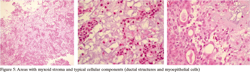
Histopathological examination (Figure 5) revealed a lacrimal gland pleomorphic adenoma, with myoepithelial cell preponderance (myxoid areas).
DISCUSSION
Tumors of the lacrimal gland are a rare condition in clinical practice, constituting 7-9% of all orbital tumors(1,2). Among all of them, the most common epithelial tumor is the lacrimal gland pleomorphic adenoma (LGPA), accounting for more than half of the epithelial forms(3), 0.6% of all orbital cases of tumors(5) and 12% of all lesions of the lacrimal gland(4).
LGPA is a benign indolent tumor, consisting of a very firm mass that leads to compression atrophy of the normal gland, displacement of residual lacrimal tissues, and is surrounded by a 'pseudocapsule' into which small sprouts of adenoma may projected(6). Most cases (90%) involve the orbital lobe of the lacrimal gland(2,5,6).
LGPA is most frequent in adults(6) in the third and fourth decades(2-4) (mean age: 41 years)(1), with no gender preponderance(1,2). The clinical presentation is usually characterized by a painless palpable mass(1,4) in the upper external quadrant of the orbit(2,8), with slow growth and inferonasal displacement of the globe(1,2,4). There may also be an increase in lacrimation and intrabulbar pressure(8), visual impairment and diplopia(2-4). Malignancy is suspected when there is a fast onset of symptoms, pain, and radiographic evidence of bone destruction(1), as in our case.
Radiological investigation may be done either by CT or MRI, as in our case. Both are similar in terms of providing information on anatomic extent, configuration, margins, and angulation features of a lacrimal gland fossa mass. However, CT provides more details about bone destruction and presence of calcification, while MRI provides better internal tissue features and intracranial extension(3,9).
On MRI, pleomorphic adenoma appears as an isointense lesion with regular margins and angles, when comparing with extraocular muscle and cerebral gray matter on T1-weighted images and heterogeneously hyperintense on T2-weighted images, due to higher water content, with moderate contrast enhancement after gadolinium-DTPA injection(9). Although not usual, there may be intratumoral hemorrhage(7), bone destruction(4,9) and calcification(4,9,10). Intracranial extension, as happened in our case, is very rare.
Although the tumor presents characteristic clinical and radiological features, which usually allows preoperative diagnosis(4,5,11), the definitive diagnosis is based on the histopathological examination(11), which shows cords of well-differentiated epithelial tubules derived from the ducts of the lacrimal gland, within loose myxomatous connective tissue(11). In our case, due to the clinical features and the extension of the tumor, a malignant tumor (epithelial carcinoma) would be the most likely diagnosis, however, an extensive histopathological examination was performed and no features of malignancy was found, confirming the diagnosis of LGPA.
Differential diagnosis includes lymphoma, chronic dacryoadenitis, Sjogren's syndrome, adenoid cystic carcinoma, granulomatous dacryoadenitis (sarcoidosis), benign lymphoid hyperplasia, cavernous hemangioma(5), intralacrimal schwannoma and hemangiopericytoma(6).
The treatment is the complete excision of the tumor and adjacent tissues, usually by lateral orbitotomy. It is believed that preoperative biopsy and incomplete resection could lead to the tumor recurrence (even after years), as well as to malignant transformation(2-7).
Complications following surgery include orbital hemorrhage, edema, optic nerve compression, orbital infection, lateral gaze palsy(2), dry eye, ptosis, lid retraction and transient diplopia(5).
The prognosis is good when the lesion is completely excised with an intact capsule(3,4). A recurrence rate of 3% within five years has been reported in complete excisions and a recurrence rate of 32% over 15 years in incomplete ones(2,3,7). It is estimated that 10% of pleomorphic adenomas undergo malignant change within 20 years after first treatment and 20% by the end of 30 years. Malignant transformation of a benign pleomorphic adenoma into a squamous cell carcinoma has also been reported 19 years after the initial operation(3).
We consider our case unusual due to its presentation at an older age, with bone destruction, invasion of the extraocular muscles and intracranial extension, which are rare features in cases of LGPA. In other words: it seems to be a malignant tumor, but it is not.
Recebido para publicação em 9/1/2013
Aceito para publicação em 17/6/2013
The authors declare no conflicts of interest
Instituição onde o trabalho foi realizado Hospital de Clínicas, Universidade Estadual de Campinas (UNICAMP) - Campinas, (SP), Brazil
- 1. Santos RR, Damasceno RW, de Pontes FS, Cursino SR, Nishiwaki-Dantas MC, Vital Filho J, et al. Ten-year follow-up of a case series of primary epithelial neoplasms of the lacrimal gland: clinical features, surgical treatment and histopathological findings. Arq Bras Oftalmol. 2010;73(1):33-9.
- 2. Halli RC, Mishra S, Kini YK, Kharkar VR, Hebbale MA. Modified lateral orbitotomy approach: a novel technique in the management of lacrimal gland tumors. J Craniofac Surg. 2011;22(3): 1035-8.
- 3. Chandrasekhar J, Farr DR, Whear NM. Pleomorphic adenoma of the lacrimal gland: case report. Br J Oral Maxillofac Surg. 2001;39(5):390-3.
- 4. Friedhofer H, Mendonça FP, Salles AG, Ferreira MC. Adenoma pleomórfico de glândula lacrimal - Relato de caso. Rev Soc Bras Cir Plast. 1997;12(3):69-74.
- 5. Prabhakaran VC, Cannon PS, McNab A, Davis G, O'Donnell B, Dolman PJ, et al. Lesions mimicking lacrimal gland pleomorphic adenoma. Br J Ophthalmol. 2010;94(11):1509-12.
- 6. Rose GE. To crash or not to crash? Probability in the management of benign lacrimal gland tumours. Eye (Lond). 2009;23(8): 1625-8.
- 7. Miyazaki T, Yamasaki T, Moritake K, Matsumoto Y, Akiyama Y, Nagai H, et al. Unusual progression of pleomorphic adenoma of the lacrimal gland: case report. Neurol Med Chir (Tokyo). 2005;45(8):407-10.
- 8. Gupta S, Garg S, Singh S, Hasija S, Chaudhary M. Pleomorphic adenoma of lacrimal gland in a 5-year-old child: Diagnosed on aspiration cytology. Diagn Cytopathol. 2013;41(6):565-6.
- 9. Gündüz K, Shields CL, Günalp I, Shields JA. Magnetic resonance imaging of unilateral lacrimal gland lesions. Graefes Arch Clin Exp Ophthalmol. 2003;241(11):907-13.
- 10. McNab AA, Satchi K. Recurrent lacrimal gland pleomorphic adenoma: clinical and computed tomography features. Ophthalmology. 2011;118(10):2088-92.
- 11. Ostrosky A, Klurfan FJ, Gonzalez MJ, Camaly D, Villa D. Pleomorphic adenoma of the lacrimal gland. Case report. Med Oral Patol Oral Cir Bucal 2005;10(1):88-9; 86-8.
Publication Dates
-
Publication in this collection
11 Nov 2013 -
Date of issue
Oct 2013
History
-
Received
09 Jan 2013 -
Accepted
17 June 2013