Abstract
Anaplasma marginale is an obligate intracellular Gram-negative bacterium found in ruminants’ erythrocytes and is the etiological agent of bovine anaplasmosis. The bacterium’s genetic diversity has been characterized based on sequences of major surface proteins (MSPs), such as MSP1α. The aim of the present study was to investigate the genetic diversity of A. marginale in cattle in the state of Maranhão, northeastern Brazil. To this end, 343 blood samples were harvested and subjected to iELISA assays using the recombinant surface protein MSP5. Out of 343 blood samples, 235 (68.5%) were randomly chosen and submitted to DNA extraction, qPCR and conventional PCR targeting the msp1α gene to determine amino acid sequences and classify the genotypes. The iELISA results showed 81.34% seropositivity (279/343), whereas qPCR revealed 224 positive samples (95.32%). Among these qPCR-positive samples, 67.4% (151/224) were also positive in the cPCR. Among the 50 obtained sequences, 21 strains had not been previously reported. Regarding the genotypes, H (26/50) and E (18/50) were identified most often, while genotypes F and C were only identified twice each and B and G once each. In conclusion, high prevalence and genetic diversity for A. marginale were observed in dairy cattle herds in the state of Maranhão.
Keywords:
Bovine anaplasmosis; tandem-repeats; MSP1α; serology
Resumo
Anaplasma marginale é uma bactéria Gram-negativa intracelular obrigatória de eritrócitos de ruminantes e responsável pela anaplasmose bovina. A diversidade genética de A. marginale tem sido caracterizada com base nas sequências das principais proteínas de superfície (MSPs), como a MSP1α. O objetivo deste estudo foi investigar a diversidade genética de A. marginale em bovinos no estado do Maranhão, Nordeste do Brasil. Dessa forma, 343 amostras de sangue foram submetidas ao ensaio iELISA, utilizando-se a proteína recombinante MSP5. Das 343 amostras de sangue, 235 (68,5%) foram escolhidas aleatoriamente e submetidas à extração de DNA, qPCR e PCR convencional para gene msp1α, para determinação das sequências de aminoácidos e classificação dos genótipos. Os resultados do iELISA mostraram 81,34% de soropositividade (279/343), enquanto qPCR revelou 224 amostras positivas (95,32%). Dentre estas na qPCR, 67,4% (151/224) mostraram-se positivas no PCR convencional. Das 50 sequências obtidas, 21 cepas não haviam sido relatadas anteriormente. Em relação aos genótipos, H (26/50) e E (18/50) foram os mais frequentes, enquanto os genótipos F e C foram identificados apenas duas vezes cada, e B e G uma vez cada. Em conclusão, alta prevalência e marcante diversidade genética de A. marginale foram observadas em rebanhos leiteiros no estado do Maranhão.
Palavras-chave:
Anaplasmose bovina; tandem-repeats; MSP1α; sorologia
Introduction
Anaplasma marginale is an obligate intracellular Gram-negative alpha-proteobacterium of the Anaplasmataceae family that parasites erythrocytes of ruminants. Because it is the etiological agent of bovine anaplasmosis, A. marginale stands out as one of the major causes of diminished bovine beef and milk production. It is also associated with high herd mortality. This bacterium parasites bovines in tropical, subtropical and temperate regions (Kocan et al., 2010; Aubry & Geale, 2011). In Brazil, bovine anaplasmosis is considered endemic (Pohl et al., 2013). A. marginale is transmitted biologically by vector ticks of the species Rhipicephalus (Boophilus) microplus and also mechanically by both hematophagous flies and contaminated fomites, may pathogen establish a persistent infection in cattle (Palmer et al., 2000). Persistent infection arises from the agent's ability to perform antigenic variation, resulting in structural changes, which make it difficult for the host's immune system to recognize it. Transplacental transmission has also been reported previously (Kocan et al., 2004; Silva et al., 2015, 2016).
The Médio Mearim microregion of Maranhão comprises 20 municipalities, occupying an area of 11,023 km2 with 411,884 inhabitants, with a population density of 37.4 inhab/km2. The area has an average altitude of 61 m (IBGE, 2018). The Microregion has a semi-humid climate, with two well-defined seasons, rainy (January to June) and dry (July to December), Cerrado and Amazon biomes, and average annual temperature ranging from 24 to 26°C (Neto et al., 2016). These characteristics contribute to the occurrence of a great diversity of ticks in the region, including R. microplus, which is the main vector of A. marginale (Costa et al., 2020). According to Medlock et al. (2013), climatic and demographic characteristics can influence the distribution and density of the R. microplus ticks.
Brazil, despite being considered enzootic for bovine anaplasmosis, has a variable prevalence according to the climatic conditions of each region (Dantas-Torres & Otranto, 2016). The molecular prevalence in the country ranges from 39.8% in semi-arid of Pernambuco (Santos et al., 2017) to 100% in São Paulo (Silva et al., 2016). Where there is a high prevalence of anaplasmosis, several A. marginale strains have been detected, and some animals may be infected with more than one strain (de la Fuente et al., 2004; Pohl et al., 2013; Machado et al., 2015; Silva et al., 2015).
The genetic diversity of A. marginale has been characterized based on sequencing major surface proteins (MSPs), such as MSP1α, MSP1β, MSP4 and MSP5 (Palmer et al., 1999). The last two were considered conserved and used for phylogenetic and molecular studies, along with serological diagnoses, whereas MSP1α is considered for a marker of the bacterium’s genetic diversity and strain variations (Cabezas-Cruz & de la Fuente, 2015). The MSP1α protein has a conserved C-terminal region and a variable N-terminal region, where sequence repetitions follow a pattern according to each existing strain of the bacterium (de la Fuente et al., 2001). Therefore, pattern variations regarding molecular weight and changing amino acids (de la Fuente et al., 2004, 2006) allow identification of different strains of A. marginale.
Thus, considering there are no studies reporting the occurrence of A. marginale in Maranhão, and that the Middle region of Mearim, Maranhão has characteristics that favor the spread of the tick R. microplus, the aim of the present study was to investigate the serological and molecular prevalence and genetic diversity of A. marginale in naturally infected cattle in dairy herds in six municipalities in the Médio Mearim microregion, Maranhão, northeastern Brazil.
Material and Methods
Study site and sampling
Blood samples were harvested between October 2018 and May 2019. The dairy cattle sampled belonged to Girolando herds on 37 farms located in six municipalities in the Médio Mearim microregion, in Maranhão, northeastern Brazil (Figure 1): 59 animals were sampled on farms 1 to 6 in the municipality of Lima Campos (4°31’14” S and 44°28’2” W); 51 on farms 7 to 12 in Pedreiras (4°34’28” S and 44°35’55” W); 52 on farms 13 to 18 in Trizidela do Vale (4°34’0” S and 44°37’37” W); 58 on farms 30 to 36 in Poção de Pedras (4°43’55” S and 44°53’18” W); 66 on farms 19 to 25 in Bernardo do Mearim (4°36’22” S and 44°46’29” W); and 57 on farms 26 to 30 and farm 37 in Igarapé Grande (4°33’19” S and 44°51’14” W).

Location and geographical distribution of farms where the bovines were sampled in Médio Mearim microregion, Maranhão, northeastern Brazil.
The farms contained an average herd of 40 to 60 animals, and approximately 10 animals of each were randomly collected. The microregion’s climate is hot with high relative humidity in the rainy season. Habitually there is a high flow of animals between farms located in the region, due to the replacement of dams/breeders and disposals due to age or death.
To determine the sample size, the Simple random sampling method was used, assuming an expected prevalence of 70% (Ramos et al., 2019), considering that the prevalence of A. marginale is unknown in the state of Maranhão, with an absolute precision of 5% and a 95% confidence interval as indicated by the formula below:
z = confidence coefficient (z = 1.96)
n = sample size
Py = expected prevalence (70%)
d2 = Absolute precision (5%)
For convenience, the blood samples were taken from 179 calves (89 males and 90 females between 1 and 12 months old) and 164 cows (> 2 years old), thus totaling 343 animals.
Two blood samples were drawn from the jugular vein of each animal. The first was collected in tubes with EDTA anticoagulant (ethylenediaminetetraacetic acid) in order to estimate the presence of rickettsemia by means of absolute quantification using qPCR and through obtaining msp1α amplicons using semi-nested PCR (snPCR). The second blood sample was collected in tubes without anticoagulant, to obtain serum for detecting IgG antibodies against A. marginale by means of the indirect enzyme-linked immunosorbent assay (iELISA). The study was approved by the Ethics Committee on Animal Use (CEEA) of the Veterinary School of the State University of Maranhão (UEMA), under protocol no. 017/2019 on June 25, 2019.
iELISA for detecting IgG antibodies against Anaplasma marginale
iELISA was performed to detect IgG antibodies against A. marginale, using recombinant major surface protein (rMSP5) of A. marginale (Imunodot® Diagnostics, Jaboticabal-SP, Brazil), in 343 cattle serum samples, according to the manufacturer’s instructions.
The plates used in the assay (Maxisorp®; Nunc, Thermo Scientific, Brazil) were coated with recombinant protein MSP5 (protein concentration was adjusted to 2.5 μg/mL) in 100 μL of sodium bicarbonate buffer 0.05 M (pH 9.6) at 4°C for 18 hours. Blockade was then performed using PBS-Tween 20 (pH 7.2) plus 6% skimmed milk powder (Molico®, Nestlé, Brazil). The plates were incubated in a humid chamber at 37°C for 90 min. After three washes with PBS-Tween 20, 100 µL of the diluted positive and negative reference serum samples, as well as the test samples, were tested in a block titration at dilutions of 1: 100 in PBS-Tween 20 and added to the ELISA plate. The plates were incubated at 37°C for 60 minutes. After three washes with PBS-Tween 20 buffer, 100 µL of anti-bovine IgG antibodies (cat. no. A0705, Sigma®, St. Louis, USA) conjugated to alkaline phosphatase were added to the ELISA plate (at a dilution of 1:30,000 for cattle, in PBS-Tween 20 solution), with subsequent incubation at 37°C for 60 minutes. After three washing steps, 100 µL of the alkaline phosphatase substrate, p-nitrophenyl phosphate (Sigma®, St. Louis, USA), was diluted to 1 mg/mL in diethanolamine buffer, pH 9.8 (Sigma®, St. Louis, USA). The plates were sealed with aluminum foil and incubated at room temperature for 45 minutes.
Readings were done on a spectrophotometer (B.T.-100; Embrabio, São Paulo, Brazil), with a 405 nm filter. The discriminant absorbance value (cutoff) was determined to be 2.5 times the average absorbance value of the negative controls (Machado et al., 1997).
The negative controls used were bovine/cattle serum samples obtained from newborn animals before colostrum intake that had been found to be negative for A. marginale through qPCR and serological assays. Serum samples positive for A. marginale through serological tests and qPCR were obtained from naturally infected animals. These serum samples had been stored in the Animal Serum Bank of the Laboratory of Immunoparasitology of the Department of Pathology, Theriogenology and One Health, UNESP Jaboticabal, SP, Brazil.
DNA extraction from blood samples
Out of 343 animals, 235 blood samples were randomly chosen and submitted to molecular analysis. DNA from 235 bovine blood samples was extracted using the InstaGene Matrix kit (Biorad™), following the manufacturer's recommendations. The DNA concentration obtained from each sample, along with their purity ratios, were measured using a spectrophotometer (NanoDrop; Thermo Scientific). The DNA samples for further PCR were stored at -20ºC.
Endogenous control PCR
To avoid false-negative results caused by the presence of inhibitors, and to check for amplifiable DNA, the DNA samples were subjected to a conventional polymerase chain reaction (cPCR) to amplify the endogenous glyceraldehyde-3-phosphate dehydrogenase gene (gapdh) of mammals, following the established protocol (Birkenheuer et al., 2003).
qPCR targeting the Anaplasma marginale msp1β gene
The samples that were positive for the gapdh gene through conventional PCR were subjected to a quantitative real-time PCR assay (qPCR) to detect the A. marginale msp1β gene (95 bp), following the protocol previously described by Carelli et al. (2007). The amplification reactions were conducted in a CFX96® thermal cycler (BioRad, Hercules, CA, United States) using the primers AM-For (5’-TTGGCAAGGCAGCAGCTT-3’) and AM-Rev (5’-TTCCGCGAGCATGTGCAT-3’) and probe Am-pb (6FAM-TCGGTCTAACATCTCCAGGCTTTCAT-BHQ1). All samples were tested in triplicates. The number of DNA copies/µL was quantified using the plasmid pSMART (Integrated DNA Technologies, Coralville, Iowa, USA), which contains the target sequence for amplifying the A. marginale DNA. The serial dilutions performed made it possible to obtain standards with different concentrations of plasmid DNA containing the target sequence (2.0 x 10⁷ to 2.0 x 100 copies/µL), determined as (X g/µL DNA/[plasmid size (bp) x 660]) x 6.022 x 10²³ x copies of the plasmid/µL. Ultrapure sterile water (Qiagen®, Madison, United States) and A. marginale DNA (Silva et al., 2015) were used as negative and positive controls, respectively, in all (q)PCR assay.
snPCR targeting the A. marginale msp1α gene
The samples that were positive for the A. marginale msp1β gene through qPCR were further subjected to snPCR targeting the msp1α gene, following the protocol previously described by Castañeda-Ortiz et al. (2015). All products from the PCR assays underwent horizontal electrophoresis and the results were visualized and analyzed using an ultraviolet light transilluminator coupled to computer software for image analysis (Chemi-Doc MP Imaging System, Bio-Rad®).
Purifying and sequencing the amplified products
The snPCR products based on the msp1α gene were purified using the ExoSAP-IT™ PCR product cleanup reagent kit (Thermo Scientific, San Jose, CA, USA), following the manufacturer's recommendations. The amplified products were sequenced using an automated technique based on the dideoxynucleotide chain termination method (Sanger et al., 1977) in an ABI PRISM 3700 DNA Analyzer sequencer (Applied Biosystem, Foster City, CA, USA). The amplimers were sequenced at the Center for Biological Resources and Genomic Biology (CREBIO) at FCAV/UNESP.
msp1α sequence analysis
The nucleotide sequences were inserted in the Phred Phrap software (Ewing et al., 1998) to screen and evaluate their quality, on electropherograms. Bases with Phred quality above 20 were considered reliable. Consensus sequences were also generated in the same software. The identities of the nucleotides obtained were compared with those in sequences deposited in GenBank (https://www.ncbi.nlm.nih.gov/genbank/) (Benson et al., 2002), using the BLASTn software (Altschul et al., 1990).
Classifying A. marginale strains and assessing genetic diversity
The nucleotide sequences of the present study and sequences previously deposited in GenBank were subject to strains classification according to Cabezas-Cruz et al. (2013), including the microsatellite genotypes and the composition of tandem repeats (TR). The 5′-UTR microsatellite is positioned between the putative Shine-Dalgarno (SD) sequence and the translation initiation codon (ATG). The structure of microsatelites was determined by GTAGG (G/A TTT)m (GT)n T ATG (Estrada-Peña et al., 2009). The analysis of the genotypes was performed according to the nomenclature proposed by de la Fuente et al. (2007). The SD-ATG distance was calculated according to the formula (4 x m) + (2 x n) +1 previously described (Estrada-Peña et al., 2009).
Using the Repeat Analyzer software, we performed the identification and analysis of tandem repeats of A. marginale msp1α (Catanese et al., 2016). Genetic diversity was calculated by metric indices that measure the percentage of single repeats in a region as well as the regularity with which repeats are distributed. Additionally, the software was used to calculate the frequency of each short-sequence repeats (SSRs) in the site under study (by the number of genotypes) and to list the sequences that were exclusive to the area under study.
Finally, the genetic diversity was calculated using the Repeat Analyzer software and was divided into two categories. The first category of metrics (GDM1) measures the amount of unique repeats in a region; while the second (GDM2) measures how uniformly the repeat occurrences in a region are distributed. GDM1 and GDM2 were presented in two variants, local and global, depending on whether the metric calculation was an average of the values of each genotype or of its region, respectively (Catanese et al., 2016). The expected values range from 0 to 1.
Statistical analysis
We used the statistical test Chi-Square (χ2) to distribute the prevalence of the disease by age group and by sampled municipalities. All statistical analyses were performed using the statistical program Graphpad Prism. 8 – Windows, with a confidence interval (CI) of 95%.
Results
Anaplasma marginale seropositivity via iELISA
The iELISA serological test using the recombinant protein MSP5 revealed the presence of IgG antibodies against A. marginale in 81.34% (279/343) of the bovine serum samples. Table 1 shows the percentage of seropositive animals in each municipality studied in the Médio Mearim macroregion, Maranhão, northeastern Brazil. Of the 279 seropositive animals, 59.50% were cows and 40.50% calves. In the prevalence of seropositivity analysis by animal age, the cows presented more seropositive animals (92.74%) when compared to the calves (68.90%), but there was no statistical difference (P = 0.15; X2 = 32.04).
Distribution of animals positive for A. marginale using the iELISA and qPCR assays in the Médio Mearim macroregion, MA.
qPCR assays for the A. marginale msp1β gene
All the 235 DNA samples were positive for the endogenous mammalian gene (gapdh) in the cPCR assay, with an average DNA concentration of 11.1 ng/µl. The 260/280 and 260/230 parameters exhibited means of 1.08 and 0.15, respectively. The qPCR assays targeting the msp1β gene indicated that 224 samples (95.32%) were positive (Table 1), with 97.12% (101/104) from calves and 93.89% (123/131) from cows (P = 0.24; X2 = 1.34). The quantity of copies of a fragment of the msp1β/μL gene ranged from 1.07 x 101 to 9.46 x 10⁹ copies, with an average of 9.72 x 10⁷ in calves and 3.19 x 10⁶ in cows. The qPCR efficiency ranged from 91.6% to 108.6%; the coefficient of determination ranged from 0.952 to 0.99; the slope ranged from -3.13 to -2.86; and the y-axis intercept ranged from 37.13 to 43.42.
cPCR for the A. marginale based on msp1α gene and BLASTn analysis
In the qPCR assays, among the 224 samples that were positive for the msp1β gene, 67.4% (151/224) were also positive for the msp1α gene fragment. Among the positive samples, it was possible to obtain 50 sequences of quality regarding the msp1α gene, through quality analysis carried out in the Phred Phrap software. The BLASTn analysis on the sequences obtained revealed percentage similarities ranging from 95% to 100%, in relation to A. marginale sequences previously deposited in the GenBank, with coverage of between 96% and 100%. The sequences of A. marginale generated in this study were deposited in the GenBank database under accession numbers: OL629247-OL629255.
Genetic diversity analysis of A. marginale based on msp1α gene
The RepeatAnalyzer software, which was used for alphanumeric genotypic classification and strain identification, detected 50 distinct strains. Among these, 21 had not previously been reported in the literature. These 21 strains are highlighted in red in Table 2.
Detected strains of the A. marginale msp1α gene, their genotypes, absolute quantification, and identification of positive animals (n = 50) sampled in Maranhão, northeastern Brazil.
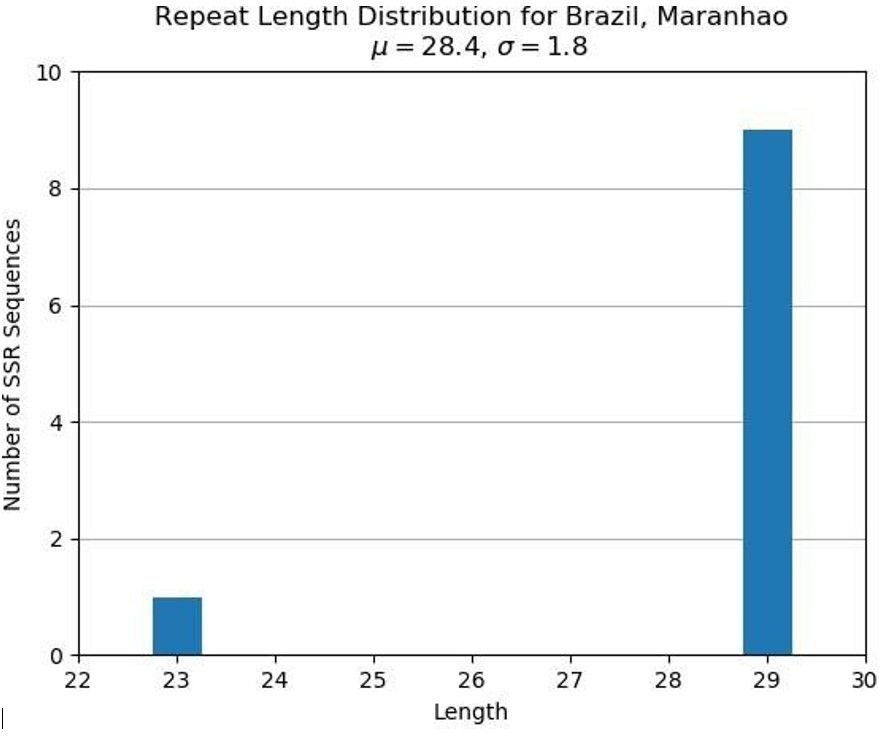
The metric indexes of A. marginale genetic diversity obtained from the dairy cattle in the Médio Mearim Region, Maranhão, were determined as GDM1-local (0.81) and GDM1-global (0.312). GDM1-local was considered high, given that this index is measured on a scale from 0 to 1. This indicates that the repetition (SSRs) was quite diverse among each other in the region studied. However, the GDM2-local (0.07) and GDM2-global (0.057) indices were low, which indicated that the SSRs were present in equal amounts, i.e. they occurred approximately the same number of times, with uniform distribution among the animals. The low GDM2 values indicated that the SSRs were sparsely dispersed. The metric indexes of genetic diversity in this study and in other Brazilian regions are shown in Table 3.
Metric indexes of genetic diversity of the A. marginale msp1α gene found in dairy cattle from different municipalities of the Médio Mearim microregion, in Maranhão, northeastern Brazil, compared to those found in other Brazilian states (São Paulo, Minas Gerais, Rio de Janeiro, Pará (buffaloes), Goiás, and Mato Grosso do Sul), as well as the values for Brazil and the world, according to the Repeat Analyzer software.
Regarding the frequencies of strains, TR Q, 15 and EV8 occurred only once; 18 and 22-2, twice; 27, τ and β, three times; α four times; and 13 five times (Figure 2). Figure 3 shows the relationship between the number of TR and the number of amino acids. The correlation between the number of genotypes and the number of SSRs found is shown in Figure 4.

Frequency of each tandem repetition found in the strains of the A. marginale msp1α gene in dairy cattle in different municipalities in the Médio Mearim microregion of Maranhão, northeastern Brazil.

Number and size (number of amino acids) of tandem repeats found in strains of the A. marginale msp1α gene in dairy cattle in different municipalities in the Médio Mearim microregion of Maranhão, northeastern Brazil.

The number of genotypes and the number of tandem repeats determined for A. marginale strains found in dairy cattle in different municipalities in the Médio Mearim microregion of Maranhão, northeastern Brazil.
The most common genotypes in the region studied were H (26/50) and E (18/50), followed far behind by F (2/50) and C (2/50). Genotypes B and G were found only once each. Figure 5 shows the distribution of the most common genotypes according to the municipality where the samples originated from.

Distribution of the most common identified genotypes according to the municipality where the samples originated from.
Discussion
Herein, the prevalence and the genetic diversity of A. marginale was assessed in northeastern Brazil. In the current study, iELISA using the recombinant protein MSP5 revealed high seropositivity rates (81.34% [279/343]) for A. marginale among the animals sampled. The high titer of IgG antibodies against A. marginale detected in the present study is corroborated by previous studies carried out in Brazil (72.2% [289/400] Ramos et al., 2019 and 91.25% [73/80] Garcia et al., 2021). Similar to seroprevalence results, the molecular prevalence found (95.32% [224/235]) is corroborated by data previously reported in different Brazilian states, such as Minas Gerais (70% - 66/94 [Pohl et al., 2013), São Paulo (94% - 47/50 [Machado et al., 2015]) and Santa Catarina (79% - 248/311 [Casa et al., 2020]), emphasizing the endemic nature of this agent in Brazil.
The prevalence of A. marginale has been associated with different risk factors i.e., cattle breeds, management system, climate, presence of ticks, production type and others (Kocan et al., 2010; Jaimes-Dueñez et al., 2018; Ola-Fadunsin et al., 2018). The climate in the Médio Mearim microregion where the present study was carried out is hot with high relative humidity in the rainy season. These environmental conditions are favorable for ticks and blood-sucking flies development and, therefore, biological and mechanical transmission of A. marginale. During sample collection, high infestation by ticks and the presence of blood-sucking insects (Haematobia irritans, Stomoxys calcitrans and tabanids) was observed (data not shown). Thus, the high prevalence found may be partially associated to presence of arthropod vectors. However, future studies are much needed to assess the epidemiological factors – i.g., iatrogenic transmission and animal trade – coupled to the high prevalence herein found.
The high seroprevalence for A. marginale in cows (92.74%) observed in this study can be also partially explained by the time these animals remain in the herd exposed to infection (Ramos et al., 2019). In calves, it is possible that immunological immaturity is related to a lower rate of seropositivity, as this age group had high levels of parasitemia (9.72 x 10⁷). Other possible causes are failure to transfer antibodies from the mother, the natural decline of antibodies during the first months of life, high infestations by ticks and flies and also transplacental transmission (Silva et al., 2015).
High diversity was observed in the microsatellite analyses and tandem repeats among A. marginale sequences obtained from sampled animals in Maranhão, northeastern Brazil. Out of 50 strains identified, 21 had never been reported in the literature, according to the RepeatAnalyzer software. The high genetic diversity of MSP1a of A. marginale seems to occur in tropical regions, as evidenced in previous studies carried out in Brazil (Machado et al., 2015; Ramos et al., 2019; Bahia et al., 2021). In addition to impacting the prevalence of bovine anaplasmosis, the presence of vectors may have direct effect on the diversity of A. marginale (Bahia et al., 2021), mainly in endemic areas (Estrada-Peña et al., 2009). Therefore, the high genetic diversity observed in the current study could be attributed to the presence of high infestation by arthropod vectors observed during the blood sampling. However, the trade of infected animals as well as herds infected for long periods may also affect the A. marginale diversity (de la Fuente et al., 2007). Thus, the factors associated with genetic diversity herein observed must be further evaluated.
Among the A. marginale strains found circulating in the sampled animals, the α-β3-Γ strain has been associated with acute anaplasmosis and high mortality in dairy herd calves on a farm located in the state of São Paulo, southeastern Brazil (Silva et al., 2016). In addition, the same strain was predominantly associated with mortality of calves, heifers, and lactating cows in Mambaí, Goiás state, central-western, Brazil (Machado et al., 2015). This A. marginale strain seems to be highly transmissible and has been previously isolated from cattle in Mexico (Almazán et al., 2008) and Argentina (Ruybal et al., 2009). Moreover, according to Cabezas-Cruz et al. (2013), this strain constitutes one of the most common A. marginale strains worldwide.
The obtained strain τ–102–15 has also been previously recorded by Silva et al. (2015) in Rio de Janeiro, southeastern Brazil. The authors suggested the probability of transplacental transmission for this strain since three animals were born infected with A. marginale presenting the MSP1a tandem repeat τ–102–15.
The genetic diversity recorded also reflected in the A. marginale genotypes observed in cattle herds from Médio Mearim, according to the classification described by Estrada-Peña et al. (2009). In the present study, H was the most frequent genotype, but genotypes E, F, C, B, and G were also found. In contrast, previous studies performed in Brazil have reported E as the most frequent genotype (Silva et al., 2016; Ramos et al., 2019; Bahia et al., 2021). According to Estrada-Peña et al. (2009), the genotype H was present across different ecoregion clusters (ecoregion clusters 2 and 3). Usually, this A. marginale genotype is related to regions where R. microplus ticks are common or in sites where R. microplus has been prevalent in the past but has been eradicated (Estrada-Peña et al., 2009).
Regarding GDM1 indexes of diversity by the RepeatAnalyzer, high genetic diversity was observed among the A. marginale genotypes analyzed, as well as in the region where the current study was performed, suggesting the prevalence of new A. marginale genotypes in Brazil. Additionally, the values of the GDM2 indices obtained suggest a low dispersion rate of these new A. marginale genotype. Similar fidings has been recorded in Brazil (Ramos et al., 2019).
Conclusions
Cattle infection with A. marginale was common in all the investigated sites from Maranhão, northeastern Brazil. High genetic diversity of A. marginale was observed in these dairy herds with 21 new strains being described and the H genotype predominating.
Acknowledgements
The authors are thankful to Imunodot Diagnosticos (Jaboticabal, SP, Brasil). This work was suppported by Fundação de Amparo à Pesquisa e ao Desenvolvimento Tecnológico do Maranhão- FAPEMA (UNIVERSAL -00749/19 FAPEMA). MRA and RZM are fellowship researchers funded by the National Council for Scientific and Technological Development (Conselho Nacional de Desenvolvimento Científico e Tecnólogico, CNPq), through grant award numbers 302420/2017-7, 306326/2014-0, respectively.
References
-
Almazán C, Medrano C, Ortiz M, de la Fuente J. Genetic diversity of Anaplasma marginale strains from an outbreak of bovine anaplasmosis in an endemic area. Vet Parasitol 2008; 158(1-2): 103-109. http://dx.doi.org/10.1016/j.vetpar.2008.08.015 PMid:18842342.
» http://dx.doi.org/10.1016/j.vetpar.2008.08.015 -
Altschul SF, Gish W, Miller W, Myers EW, Lipman DJ. Basic local alignment search tool. J Mol Biol 1990; 215(3): 403-410. http://dx.doi.org/10.1016/S0022-2836(05)80360-2 PMid:2231712.
» http://dx.doi.org/10.1016/S0022-2836(05)80360-2 -
Aubry P, Geale DW. A review of bovine anaplasmosis. Transbound Emerg Dis 2011; 58(1): 1-30. http://dx.doi.org/10.1111/j.1865-1682.2010.01173.x PMid:21040509.
» http://dx.doi.org/10.1111/j.1865-1682.2010.01173.x -
Bahia M, Silva LT, Silva BM, Cordeiro MD, Guterres A, Silva CB, et al. Genetic diversity of Anaplasma marginale in calves with anaplasmosis on farms in Minas Gerais, Brazil. Ticks Tick Borne Dis 2021; 12(1): 101552. http://dx.doi.org/10.1016/j.ttbdis.2020.101552 PMid:33010632.
» http://dx.doi.org/10.1016/j.ttbdis.2020.101552 -
Benson DA, Karsch-Mizrachi I, Lipman DJ, Ostell J, Rapp BA, Wheeler DL. GenBank. Nucleic Acids Res 2002; 30(1): 17-20. http://dx.doi.org/10.1093/nar/30.1.17 PMid:11752243.
» http://dx.doi.org/10.1093/nar/30.1.17 -
Birkenheuer AJ, Levy MG, Breitschwerdt EB. Development and evaluation of a seminested PCR for detection and differentiation of Babesia gibsoni (Asian genotype) and B. canis DNA in canine blood samples. J Clin Microbiol 2003; 41(9): 4172-4177. http://dx.doi.org/10.1128/JCM.41.9.4172-4177.2003 PMid:12958243.
» http://dx.doi.org/10.1128/JCM.41.9.4172-4177.2003 -
Cabezas-Cruz A, de la Fuente J. Anaplasma marginale major surface protein 1a: a marker of strain diversity with implications for control of bovine anaplasmosis. Ticks Tick Borne Dis 2015; 6(3): 205-210. http://dx.doi.org/10.1016/j.ttbdis.2015.03.007 PMid:25802034.
» http://dx.doi.org/10.1016/j.ttbdis.2015.03.007 -
Cabezas-Cruz A, Passos LMF, Lis K, Kenneil R, Valdés JJ, Ferrolho J, et al. Functional and immunological relevance of Anaplasma marginale major surface protein 1a sequence and structural analysis. PLoS One 2013; 8(6): e65243. http://dx.doi.org/10.1371/journal.pone.0065243 PMid:23776456.
» http://dx.doi.org/10.1371/journal.pone.0065243 -
Carelli G, Decaro N, Lorusso A, Elia G, Lorusso E, Mari V, et al. Detection and quantification of Anaplasma marginale DNA in blood samples of cattle by real-time PCR. Vet Microbiol 2007; 124(1-2): 107-114. http://dx.doi.org/10.1016/j.vetmic.2007.03.022 PMid:17466470.
» http://dx.doi.org/10.1016/j.vetmic.2007.03.022 -
Casa MS, Vettori JM, Souza KM, Miletti LC, Vogel CIG, Lima ALF, et al. High prevalence of Anaplasma marginale in the Crioula Lageana cattle. J Infect Dev Ctries 2020; 14(6): 623-630. http://dx.doi.org/10.3855/jidc.11691 PMid:32683353.
» http://dx.doi.org/10.3855/jidc.11691 -
Castañeda-Ortiz EJ, Ueti MW, Camacho-Nuez M, Mosqueda JJ, Mousel MR, Johnson WC, et al. Correction: association of Anaplasma marginale strain superinfection with infection prevalence within tropical regions. PLoS One 2015; 10(5): e0129415. http://dx.doi.org/10.1371/journal.pone.0129415 PMid:26020787.
» http://dx.doi.org/10.1371/journal.pone.0129415 -
Catanese HN, Brayton KA, Gebremedhin AH. RepeatAnalyzer: a tool for analysing and managing short-sequence repeat data. BMC Genomics 2016; 17(1): 422. http://dx.doi.org/10.1186/s12864-016-2686-2 PMid:27260942.
» http://dx.doi.org/10.1186/s12864-016-2686-2 -
Costa FB, Martins TF, Muñoz-Leal S, de Azevedo Serpa MC, Ogrzewalska M, Luz HR, et al. Retrospective and new records of ticks (Acari: Argasidae, Ixodidae) from the state of Maranhão, an Amazon-Cerrado transition area of Brazil. Vet Parasitol Reg Stud Reports 2020; 21: 100413. http://dx.doi.org/10.1016/j.vprsr.2020.100413 PMid:32862893.
» http://dx.doi.org/10.1016/j.vprsr.2020.100413 - Dantas-Torres F, Otranto D. Anaplasmosis. In: Marcondes CB, editor. Arthropod borne diseases . Cham: Springer; 2016. p. 215-222.
-
Estrada-Peña A, Naranjo V, Acevedo-Whitehouse K, Mangold AJ, Kocan KM, de la Fuente J. Phylogeographic analysis reveals association of tick-borne pathogen, Anaplasma marginale, MSP1a sequences with ecological traits affecting tick vector performance. BMC Biol 2009; 7(1): 57. http://dx.doi.org/10.1186/1741-7007-7-57 PMid:19723295.
» http://dx.doi.org/10.1186/1741-7007-7-57 -
Ewing CA, Hillier L, Wendl MC, Green P. Base-calling of automated sequencer traces using phred. I. Accuracy assessment. Genome Res 1998; 8(3): 175-185. http://dx.doi.org/10.1101/gr.8.3.175 PMid:9521921.
» http://dx.doi.org/10.1101/gr.8.3.175 -
de la Fuente J, Almazán C, Blouin EF, Naranjo V, Kocan KM. Reduction of tick infections with Anaplasma marginale and A. phagocytophilum by targeting the tick protective antigen subolesin. Parasitol Res 2006; 100(1): 85-91. http://dx.doi.org/10.1007/s00436-006-0244-6 PMid:16816958.
» http://dx.doi.org/10.1007/s00436-006-0244-6 -
de la Fuente J, Garcia-Garcia JC, Blouin EF, Rodríguez SD, García MA, Kocan KM. Evolution and function of tandem repeats in the major surface protein 1a of the ehrlichial pathogen Anaplasma marginale. Anim Health Res Rev 2001; 2(2): 163-173. http://dx.doi.org/10.1079/AHRR200132 PMid:11831437.
» http://dx.doi.org/10.1079/AHRR200132 -
de la Fuente J, Passos LMF, Van Den Bussche RA, Ribeiro MFB, Facury-Filho EJ, Kocan KM. Genetic diversity and molecular phylogeny of Anaplasma marginale isolates from Minas Gerais, Brazil. Vet Parasitol 2004; 121(3-4): 307-316. http://dx.doi.org/10.1016/j.vetpar.2004.02.021 PMid:15135871.
» http://dx.doi.org/10.1016/j.vetpar.2004.02.021 -
de la Fuente J, Ruybal P, Mtshali MS, Naranjo V, Shuqing L, Mangold AJ, et al. Analysis of world strains of Anaplasma marginale using major surface protein 1a repeat sequences. Vet Microbiol 2007; 119(2-4): 382-390. http://dx.doi.org/10.1016/j.vetmic.2006.09.015 PMid:17084044.
» http://dx.doi.org/10.1016/j.vetmic.2006.09.015 -
Garcia AB, Jusi MMG, Freschi CR, Ramos IAS, Mendes NS, Amaral RB, et al. High genetic diversity and superinfection by Anaplasma marginale strains in naturally infected Angus beef cattle during a clinical anaplasmosis outbreak in southeastern Brazil. Ticks Tick Borne Dis 2021; 13(1): 101829. http://dx.doi.org/10.1016/j.ttbdis.2021.101829 PMid:34798528.
» http://dx.doi.org/10.1016/j.ttbdis.2021.101829 -
Instituto Brasileiro de Geografia e Estatística – IBGE. Série histórica da produção de leite no Maranhão [online]. Rio de Janeiro: IBGE; 2018 [cited 2020 Aug 25]. Available from: https://cidades.ibge.gov.br/brasil/ma/pesquisa/18/16459?tipo=grafico&indicador=16559
» https://cidades.ibge.gov.br/brasil/ma/pesquisa/18/16459?tipo=grafico&indicador=16559 -
Jaimes-Dueñez J, Triana-Chávez O, Mejía-Jaramillo AM. Genetic, host and environmental factors associated with a high prevalence of Anaplasma marginale. Ticks Tick Borne Dis 2018; 9(5): 1286-1295. http://dx.doi.org/10.1016/j.ttbdis.2018.05.009 PMid:29793771.
» http://dx.doi.org/10.1016/j.ttbdis.2018.05.009 -
Kocan KM, de la Fuente J, Blouin EF, Coetzee JF, Ewing SA. The natural history of Anaplasma marginale. Vet Parasitol 2010; 167(2-4): 95-107. http://dx.doi.org/10.1016/j.vetpar.2009.09.012 PMid:19811876.
» http://dx.doi.org/10.1016/j.vetpar.2009.09.012 -
Kocan KM, de la Fuente J, Blouin EF, Garcia-Garcia JC. Anaplasma marginale (Rickettsiales: Anaplasmataceae): recent advances in defining host-pathogen adaptations of a tick-borne rickettsia. Parasitology 2004;129(Suppl S1): S285-S300. http://dx.doi.org/10.1017/S0031182003004700 PMid:15938516.
» http://dx.doi.org/10.1017/S0031182003004700 -
Machado RZ, Montassier HJ, Pinto AA, Lemos EG, Machado MR, Valadão IF, et al. An enzyme-linked immunosorbent assay (ELISA) for the detection of antibodies against Babesia bovis in cattle. Vet Parasitol 1997; 71(1): 17-26. http://dx.doi.org/10.1016/S0304-4017(97)00003-4 PMid:9231985.
» http://dx.doi.org/10.1016/S0304-4017(97)00003-4 -
Machado RZ, Silva JB, André MR, Gonçalves LR, Matos CA, Obregón D. Outbreak of anaplasmosis associated with the presence of different Anaplasma marginale strains in dairy cattle in the states of São Paulo and Goiás, Brazil. Rev Bras Parasitol Vet 2015; 24(4): 438-446. http://dx.doi.org/10.1590/S1984-29612015078 PMid:26648009.
» http://dx.doi.org/10.1590/S1984-29612015078 -
Medlock JM, Hansford KM, Bormane A, Derdakova M, Estrada-Peña A, George JC, et al. Driving forces for changes in geographical distribution of Ixodes ricinus ticks in Europe. Parasit Vectors 2013; 6(1): 1. http://dx.doi.org/10.1186/1756-3305-6-1 PMid:23281838.
» http://dx.doi.org/10.1186/1756-3305-6-1 -
Neto CAAP, Lima GP, Chaves LPFA, Silva RA, Siqueira GM. Biogeography of the Central Mesoregion of Maranhão (Brazil). J Geospat Modelling 2016; 1(1): 33-40. http://dx.doi.org/10.22615/jgm-1.1-5812
» http://dx.doi.org/10.22615/jgm-1.1-5812 -
Ola-Fadunsin SD, Gimba FI, Abdullah DA, Sharma RSK, Abdullah FJF, Sani RA. Epidemiology and risk factors associated with Anaplasma marginale infection of cattle in Peninsular Malaysia. Parasitol Int 2018; 67(6): 659-665. http://dx.doi.org/10.1016/j.parint.2018.06.013 PMid:29960083.
» http://dx.doi.org/10.1016/j.parint.2018.06.013 -
Palmer GH, Brown WC, Rurangirwa FR. Antigenic variation in the persistence and transmission of the ehrlichia Anaplasma marginale. Microbes Infect 2000; 2(2): 167-176. http://dx.doi.org/10.1016/S1286-4579(00)00271-9 PMid:10742689.
» http://dx.doi.org/10.1016/S1286-4579(00)00271-9 -
Palmer GH, Rurangirwa FR, Kocan KM, Brown WC. Molecular basis for vaccine development against the ehrlichial pathogen Anaplasma marginale. Parasitol Today 1999; 15(7): 281-286. http://dx.doi.org/10.1016/S0169-4758(99)01469-6 PMid:10377531.
» http://dx.doi.org/10.1016/S0169-4758(99)01469-6 -
Pohl AE, Cabezas-Cruz A, Ribeiro MFB, Silveira JAG, Silaghi C, Pfister K, et al. Detection of genetic diversity of Anaplasma marginale isolates in Minas Gerais, Brazil. Rev Bras Parasitol Vet 2013; 22(1): 129-135. http://dx.doi.org/10.1590/S1984-29612013000100024 PMid:24252959.
» http://dx.doi.org/10.1590/S1984-29612013000100024 -
Ramos IAS, Herrera HM, Fernandes SJ, Amaral RB, Zanatto DCS, Silva TMV, et al. Genetic diversity of Anaplasma marginale in beef cattle in the Brazilian Pantanal. Ticks Tick Borne Dis 2019; 10(4): 805-814. http://dx.doi.org/10.1016/j.ttbdis.2019.03.015 PMid:30930114.
» http://dx.doi.org/10.1016/j.ttbdis.2019.03.015 -
Ruybal P, Moretta R, Perez A, Petrigh R, Zimmer P, Alcaraz E, et al. Genetic diversity of Anaplasma marginale in Argentina. Vet Parasitol 2009; 162(1-2): 176-180. http://dx.doi.org/10.1016/j.vetpar.2009.02.006 PMid:19285808.
» http://dx.doi.org/10.1016/j.vetpar.2009.02.006 -
Sanger F, Nicklen S, Coulson A. DNA sequencing with chain-terminating inhibitors. Proc Natl Acad Sci USA 1977; 74(12): 5463-5467. http://dx.doi.org/10.1073/pnas.74.12.5463 PMid:271968.
» http://dx.doi.org/10.1073/pnas.74.12.5463 -
Santos GB, Gomes IM, Silveira JAG, Pires LCSR, Azevedo SS, Antonelli AC, et al. Cattle tick fever in semi-arid of Pernambuco. Pesq Vet Bras 2017; 37(1): 1-7. http://dx.doi.org/10.1590/s0100-736x2017000100001
» http://dx.doi.org/10.1590/s0100-736x2017000100001 -
Silva JB, André MR, Machado RZ. Low genetic diversity of Anaplasma marginale in calves in an endemic area for bovine anaplasmosis in the state of São Paulo, Brazil. Ticks Tick Borne Dis 2016; 7(1): 20-25. http://dx.doi.org/10.1016/j.ttbdis.2015.07.018 PMid:26318264.
» http://dx.doi.org/10.1016/j.ttbdis.2015.07.018 -
Silva JB, Fonseca AH, Barbosa JD, Cabezas-Cruz A, de la Fuente J. Low genetic diversity associated with low prevalence of Anaplasma marginale in water buffaloes in Marajó Island, Brazil. Ticks and tick-borne diseases 2014; 5(6): 801-804. 10.1016/j.ttbdis.2014.06.003.
» https://doi.org/10.1016/j.ttbdis.2014.06.003 -
Silva JB, Gonçalves LR, Varani AM, André MR, Machado RZ. Genetic diversity and molecular phylogeny of Anaplasma marginale studied longitudinally under natural transmission conditions in Rio de Janeiro, Brazil. Ticks Tick Borne Dis 2015; 6(4): 499-507. http://dx.doi.org/10.1016/j.ttbdis.2015.04.002 PMid:25985719.
» http://dx.doi.org/10.1016/j.ttbdis.2015.04.002 - Thrusfield M. Veterinary epidemiology 3rd ed. Oxford: Blackwell Science Ltd.; 2006. Types of sampling; p. 230-246.