Megakaryocytes are cells responsible for the production of platelets, and present unique morphological characteristics, particularly their great size and certain singular aspects of their cytoplasm and nucleus(1,2). Thus, they are easy to identify under routine microscopic examination. Frequently cytologic and/or histologic bone marrow analysis enables the diagnosis or detection of hematologic disorders that involve megakaryocytes(3). Among these disorders myelodysplastic syndromes and myeloproliferative neoplasms stand out(4,5) (Figures 1–8).
(A) Normal megakaryocytes from bone marrow aspirate smear (cytology, Leishmann staining, X1000). (B) Normal megakaryocytes from bone marrow biopsy (histopathology, hematoxylin and eosin, X1000). It is worth noting that normal megakaryocytes present a characteristic cohese nucleus with many lobes. (C) Bone marrow immunohistochemistry, identifying megakaryocytes by positive staining for von Willebrand factor, which is present on cell surface
Monolobulated megakaryocytes (hematoxylin and eosin, X1000), characteristic of the “5q- syndrome”. This subtype of myelodysplastic syndrome is characterized by macrocytic anemia, normal or increased platelet count, deletion of the long arm of chromossome 5 as the sole cytogenetic anomaly, good prognosis and good response to lenalidomide
Bone marrow karyotype (G band), highlighting the characteristic anomaly of the 5q-syndrome (interstitial deletion of chromosome 5)
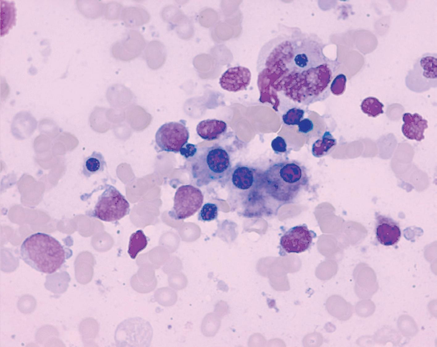
Micromegakaryocytes in bone marrow aspirate smear (Leismann staining, X1000), representing small monolobulated cells (smaller than two neutrophyls). Although common in myelodydsplastic syndromes, they have no diagnostic specificity
Abnormal multinucleated megakaryocytes presenting karyolysis in bone marrow aspirate smear (Leishmann staining, X1000). Although common in myelodydsplastic syndromes, they have no diagnostic specificity
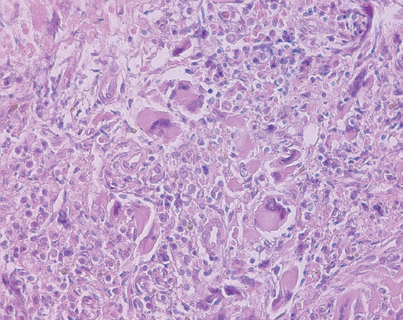
Megakaryocyte in myeloproliferative neoplasm, characterized by cluster formation. The morphologic aspect does not allow identifying the subtype of myeloproliferation, which could occur in chronic myeloid leukemia (CML), myelofibrosis (cellular phase), polycythemia vera, or essential thrombocythemia. Correct diagnosis depends on molecular studies to identify the BCR-ABL rearrangement (diagnosis of CML) or a JAK-2 mutation (myelofibrosis, polycythemia vera or essential thrombocythemia)
REFERENCES
- 1 Bluteau D, Lordier L, Di Stefano A, Chang Y, Raslova H, Debili N, et al. Regulation of megakaryocyte maturation and platelet formation. Thromb Haemost. 2009;7 Suppl 1:227-34.
- 2 Thon JN, Italiano JE. Platelet formation. Semin Hematol. 2010;47(3):220-6. Review.
- 3 Tang G, Wang SA, Menon M, Dresser K, Woda BA, Hao S. High-level CD34 expression on megakaryocytes independently predicts an adverse outcome in patients with myelodysplastic syndromes. Leuk Res. 2011 Feb 28. [Epub ahead of print]
- 4 Chauffaille MLL. Neoplasias mieloproliferativas: revisão dos critérios diagnósticos e dos aspectos clínicos. Rev Bras Hematol Hemoter. 2010;32(4):308-16.
- 5 Hall J, Foucar K. Diagnosing myelodysplastic/myeloproliferative neoplasms: laboratory testing strategies to exclude other disorders. Int J Lab Hematol. 2010;32(6 Pt 2):559-71.

 Megakaryocyte


