Abstracts
The Stroke is a neurological disorder that disables more adults. The breathing, posture and swallowing have a direct relationship in this disease, thus requiring a multidisciplinary approach. The goal of this study was to conduct a clinical evaluation integrated to analyze the relationship between these disorders. This case is a female patient aged 37 with six years of diagnosis of ischemic stroke with right hemiparesis complete predominantly crural, hypertension, dysphagia and dysarthrophonia. In history, reported symptoms related to dyspnea, pain in the lumbar spine, the presence of coughing and choking and drooling to food, especially during the speech. The patient has scoliosis, thoracic kyphosis and abdominal weakness. The evaluation showed a respiratory expiratory force below the expected range. The peak cough flow is lower than expected, demonstrating not have the strength to cough. The research revealed changes in speech increased time to eat, need to drink to help swallow solid food before any leaks, the need for multiple swallows due to the sense of stasis in laryngeal-pharyngeal cavity and decreased taste and thick and viscous saliva. Using physical and speech therapy evaluations, we concluded that a patient with stroke in chronic respiratory presents alterations that may prevent it from effectively protecting the lower airways, which, added to postural changes and swallowing show the importance of integrated assessment for future therapeutic interventions more effective.
Stroke; Deglutition; Physiotherapy; Speech, Language and Hearing Sciences; Posture; Respiration
O Acidente Vascular Cerebral (AVC) é a doença neurológica que mais incapacita os adultos. A respiração, a postura e a deglutição possuem uma relação direta nesta doença, necessitando assim de uma intervenção multidisciplinar. O objetivo do presente estudo foi realizar uma avaliação clínica integrada para analisar a relação entres estas disfunções. Trata-se de uma paciente do sexo feminino com 37 anos, com seis anos de diagnóstico de AVC isquêmico com hemiparesia completa à direita de predomínio crural, hipertensão arterial sistêmica, disfagia e disartrofonia. Na anamnese, relatou sintomas referentes à dispnéia, dores na coluna lombar, presença de tosse e engasgos à alimentação e escape anterior de saliva, principalmente durante a fala. A paciente apresenta escoliose, hipercifose torácica e fraqueza dos músculos abdominais. A avaliação respiratória evidenciou uma força expiratória abaixo do limite esperado. O pico de fluxo da tosse está abaixo do esperado, demonstrando não possuir força para tossir. A investigação fonoaudiológica evidenciou alterações no tempo aumentado para se alimentar, necessidade de ingerir líquidos para ajudar na deglutição de sólidos, eventuais escapes anteriores de alimento, necessidade de deglutições múltiplas devido à sensação de estase em cavidade laringo-faríngea e diminuição do paladar, bem como saliva grossa e viscosa. Utilizando-se das avaliações fisioterápicas e fonoaudiológicas, foi possível concluir que uma paciente com AVC na fase crônica apresenta alterações respiratórias que podem impedi-la de proteger com eficiência as vias aéreas inferiores, o que, somado às alterações posturais e na deglutição demonstram a importância da avaliação integrada para futuras intervenções terapêuticas mais eficazes.
Acidente Cerebral Vascular; Deglutição; Fisioterapia; Fonoaudiologia; Postura; Respiração
CASE REPORTS
IPhysical Therapist; Specialization Degree in Cardiorespiratory Physical Therapy from UNINOVE - Universidade Nove de Julho, São Paulo, SP; Specialization Degree in Neurological Rehabilitation from Unifesp - Universidade Federal de São Paulo, São Paulo SP, Brazil
IISpeech Therapist; Master's Student in Rehabilitation Sciences at USP - Universidade de São Paulo, Specialization Degree in Neurological Rehabilitation from Unifesp - Universidade Federal de São Paulo, São Paulo SP, Brazil; Specialization Degree in Language from USP - Universidade de São Paulo, São Paulo, SP, Brazil
IIIPhysical Therapist; Professor at Centro Universitário São Camilo and Universidade Nove de Julho; MSc in Neurosciences from Unifesp - Universidade Federal de São Paulo, São Paulo SP, Brazil
IVSpeech Therapist; PhD in Neurosciences from Unifesp - Universidade Federal de São Paulo, São Paulo, SP, Brazil
VRespiratory Physical Therapist at the Outpatient Service of the Neuromuscular Disorders Investigation Center of Unifesp - Universidade Federal de São Paulo, São Paulo, SP, Brazil
VISpeech Therapist; PhD in Neurosciences from Unifesp - Universidade Federal de São Paulo, São Paulo, SP, Brazil
VIIMD, Neurologist; PhD student at Unifesp - Universidade Federal de São Paulo, São Paulo, SP, Brasil; MSc in Vascular Neurology from Unifesp - Universidade Federal de São Paulo, São Paulo, SP, Brazil
Mailing Address
ABSTRACT
The Stroke is a neurological disorder that disables more adults. The breathing, posture and swallowing have a direct relationship in this disease, thus requiring a multidisciplinary approach. The goal of this study was to conduct a clinical evaluation integrated to analyze the relationship between these disorders. This case is a female patient aged 37 with six years of diagnosis of ischemic stroke with right hemiparesis complete predominantly crural, hypertension, dysphagia and dysarthrophonia. In history, reported symptoms related to dyspnea, pain in the lumbar spine, the presence of coughing and choking and drooling to food, especially during the speech. The patient has scoliosis, thoracic kyphosis and abdominal weakness. The evaluation showed a respiratory expiratory force below the expected range. The peak cough flow is lower than expected, demonstrating not have the strength to cough. The research revealed changes in speech increased time to eat, need to drink to help swallow solid food before any leaks, the need for multiple swallows due to the sense of stasis in laryngeal-pharyngeal cavity and decreased taste and thick and viscous saliva. Using physical and speech therapy evaluations, we concluded that a patient with stroke in chronic respiratory presents alterations that may prevent it from effectively protecting the lower airways, which, added to postural changes and swallowing show the importance of integrated assessment for future therapeutic interventions more effective.
Keywords: Stroke; Deglutition; Physiotherapy; Speech, Language and Hearing Sciences; Posture; Respiration
INTRODUCTION
Stroke is the most frequent cephalic vascular condition, whose most common type is the ischemic type1. Changes in the circulatory system are very relevant in the morbidity and mortality rates of the country, in which cerebrovascular conditions stand out. This condition particularly represents the third leading cause of death in industrialized countries, the first leading cause of disability of adults2, and, in Brazil, the first leading cause of death of adults. It is considered the most physically disabling condition1,3 and has great impact on costs of diagnosis, treatment and rehabilitation4.
The motor deficits in this condition result from damage to superior motor neurons that control muscles and, along with respiratory muscles, help enable movements of the trunk5,6.
Thus, respiratory disorders may promote significant sensorial, postural, motor and occupational changes. The shortening of inspiratory muscles reduces the elasticity and expansibility of the chest cavity. Although these muscles must contract, their motor activities are marked by frailty. Several factors may change this pattern, such as the amount of secretion, presence of bronchospasm, emotional and behavioral aspects, weight reduction and poor postures7, such as scoliosis. Therefore, they may interfere both in posture and breathing.
Neurogenic dysphagia includes changes in the swallowing function, resulting from a neurological disease with symptoms and complications deriving from sensory and motor impairment of the muscles involved in swallowing. Among these neurological disorders, stroke seems to be the most common cause of dysphagia8.
Assessment of the oropharyngeal dysphagia and the implementation of prophylactic and therapeutical measures among patients seem to be capable of reducing the rate of clinical complications associated with these changes9. This assessment can be conducted by different methods. There is no consensus about the best non-invasive clinical investigation standard, except that it should have a high level of sensitivity for identification of aspiration and it should be safe for the patient10.
Breathing and swallowing are highly coordinated processes using the same physiological structures. Changes in both processes have been recorded after stroke, among which the increased risk of aspiration when eating is of striking relevance11. In respect to posture, it is known that a great part of the problems related to oral functions occurs because of inappropriate body posture12.
Integrated physical and phonoaudiological evaluations are extremely important to allow a more thoroughly detailed investigation of posture, breathing and swallowing, leading to a more complete and integrated diagnosis, and thus enabling an interdisciplinary therapeutic approach.
The purpose of this study was to conduct an integrated clinical evaluation of the relationship between these dysfunctions.
METHOD
We report the case of a female patient, duly enrolled in the Neurovascular Diseases Center of Unifesp - Universidade Federal de São Paulo, where she undergoes periodical neurological follow-up.
Physical therapy procedures included assessment of the heart rate, breathing rate and blood pressure, in addition to assessment of chest mobility by means of palpation and measurement of chest perimeters (axillary and xyphoid process).
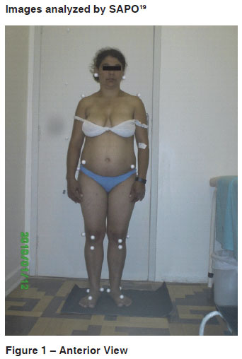
The quantitative postural evaluation used the SAPO13 protocol, which is a Postural Evaluation Software, and the respective scoring. The test required a personal computer with Windows XP and a Mitsuca DS 507IBR digital camera, 5.0 megapixels, model DSCS85. The camera was placed on a stand, 0.75 cm above and parallel to the floor, 2.35 m away from the patient. A piece of black card stock and a silver marker were used to indicate positioning of the feet and allow that the same support basis be used in all pictures. For the first picture, the plumb line had 1 m and 0.40 cm references and 25 mm body marks for calibration of the images. The patient was asked to walk on the same place and then stop as comfortably as possible. Pictures were taken from the anterior, right lateral, left lateral and posterior view, and later analyzed by the system.
The respiratory evaluation included measurement of the Maximal Inspiratory Pressure (PiMax)14 and Maximal Expiratory Pressure (PeMax)14, using a Imebras - 150 manovacuometer, as well as the Vital Capacity (V.C.)15, using the Ferraris Wright MK8 ventilometer, and the Peak Cough Flow (P.C.F.)15, using the Peak Flow Harlow Airmed CM 20 2 TT.
The BERG12 scale was used to assess balance changes, and the cognitive evaluation included the Mini-Mental Estate Exam (MMEE)16 and the clock test17.
Phonoaudiological analysis procedures were conducted based on the Phonoaudiological Investigation Protocol of the Neuromuscular Disorders Investigation Center of UNIFESP18, involving phonoaudiological history and clinical evaluation of stomatognathic system muscles, as well as voice, speech, chewing and swallowing functions.
The eating test was conducted in four stages: saliva, thin liquids, thick liquids and solids. The thin (water) and thick (yogurt) liquids were administered in amounts of 1, 3, 5 and 10 ml in a disposable syringe; the solid was a day-old French roll, as per the Phonoaudiological Investigation Protocol of the Neuromuscular Disorders Center of UNIFESP18.
This research study was approved by the Ethics Committee of Universidade Federal de São Paulo / Hospital São Paulo under record number 1831/09. The purposes of the study were communicated to the patient and caretaker through reading of the Informed Consent Form, which was signed after the patient accepted to take part in this study.
After collection of the aforementioned data, a descriptive analysis and discussion were conducted.
CASE INTRODUCTION
Patient L.S.L, 37, female, housewife, basic education incomplete, with medical diagnosis of Ischemic Stroke in the left hemisphere and physical diagnosis of complete espastic hemiparesis, right-sided, disproportional, with crural predominance, after two ischemic events of cardioembolic origin. The first was in 2001 with incomplete espastic hemiparesis, right-sided, disproportional, with crural predominance, and the second in 2002, with involvement of the right face.
In her physical and phonoaudiological history, the patient reported symptoms including dyspnea, lumbar pain, coughing and choking on food, difficulty articulating speech sounds and excessive saliva, mainly during speech; previous conditions included systemic arterial hypertension (SAH). She denied any vertigo, smoking, drinking and other addictions.
The initial evaluation showed scoliosis, thoracic hyperkyphosis and weakness of abdominal muscles, hemiparetic gait, no assistive gait device and incapacity to perform triple flexion (hip, knee and dorsiflexion), facial asymmetry with predominantly right-sided laxity, absent teeth on both dental archs, presence of fur over the tongue surface and signs of right-sided atrophy of the tongue, breathy vocal quality and imprecise articulation of speech sounds.
RESULTS
The physical clinical investigation detected: elastic hypertonia, weakness of abdominal muscles and globally on the hemiparetic side, and gait present without any assistive gait device.
The balance test using the BERG scale was satisfactory, and a score of 55 out of 56 was obtained. The only difficulty observed was for criterion "reaching forward with arms extended", for which displacement of the gravity center was required.
Cognitive evaluation through the MMEE and the clock test demonstrated a significant cognitive deficit, which may have hindered understanding of some tests; the patient, however, has good recent memory and object naming capacity. In the MMEE criteria for orientation, the patient scored 2 out of 10; for attention and calculation, 0 out of 5; in language evaluation, 6 out of 9; and for the clock evaluation, she scored one, being able to draw only the outer circle of the clock.
SAPO provides results after analyzing images entered in the four-view system, as per Figures 1, 2, 3, and 4 . We observed that the patient has anteriorization of the head, elevation of the left shoulder, flexion of the trunk, elevation of the right anterosuperior iliac spine, antepulsion, pelvic anteversion and scoliosis with left-sided convexity. The clock generated also provided information on the projection of the center of gravity (C.G.), with observation of considerable frontal displacement of the C.G and weight bearing slightly shifted to the left hemibody (Figures 1 to 4).
On the respiratory evaluation, the patient did not have any discomfort that could hinder the progress of the study. The respiratory evaluation indicated good strength of respiratory muscles, with PiMax=80 CmH2O, and lower limit at -77.94 CmH2O; expiratory force, however, was below the lower limit of 75.02 CmH2O, with PeMax =70 CmH2O. The P.C.F. was 200, which was below expected, i.e. the patient did not have enough strength to cough. The V.C. was 1.5 L, using just 47% of the expected capacity.
The phonoaudiological investigation revealed the following changes related to dysphagia: increased time to eat, the need to drink to help swallow solid and dry foods, occasional anterior food loss, the need for multiple swallows due to sensation of stasis in the laryngeal-pharyngeal cavity, decreased taste function, and increased saliva thickness and viscosity.
Evaluation of the stomatognathic system showed facial asymmetry at rest and motion, with more pronounced laxity of the right face. In respect to occlusion: absence of dental elements on the inferior (1st left molar, right canine, 1st and 3rd right molars) and the superior arch (left premolar and 1st molar, and right premolar, 1st and 2nd molar).
The lip was more horizontalized on the right face when smiling, and lip puckering showed higher strength on the left face, with grade 1 muscle strength (mild). The tongue surface is moderately covered with fur and the tip is shifted to the left; patient shows fatigue during performance of isotonic movements and there are signs of atrophy on the right side, with grade 1 (mild) muscle strength. Buccinators show grade 1 muscle strength (mild) on the left side and grade 2 (moderate) on the right side.
Analysis of the chewing function showed deficient pattern, with right-sided unilateral mastication (despite the lower strength on this side), reduced range, incomplete and slow rotatory movement, with normal cutting of the food and no changes in the chewing-swallowing coordination.
Swallowing was rated as normal for saliva and thin liquids, in the amounts of 1, 3 and 5 ml. The offer of 10 ml of liquid resulted in mildly wet voice, with no episodes of coughing or choking. For thick liquids (yogurt), there was residue on the hard and soft palate, vestibule and dorsal tongue with the amounts of 5 and 10 ml, which had not been removed by the third swallow. For solids (day-old French roll), a large amount of residue was still observed, even after the fourth swallow. The chewing-swallowing coordination was normal.
The voice quality was breathy (grade 2) and shaky (grade 1), with low maximal times for the respective age and gender (/a/ = 9 seconds (mean); /i/ = 4 seconds and /u/ = 5 seconds); during emission, there was instability (mild to moderate), mild asthenia, sonority breaks, decreased volume and intensity (moderate) and mild use of reserve air volume. Pitch was normal, loudness was reduced and resonance showed excessive use of the larynx. On the vocal resistance test, vocal quality, breathing dynamics and intensity were not sustained, and the patient abandoned before completion.
Speech showed decreased velocity; changes in prosody, with the use of short sentences and reduced emphasis; and highly impaired articulation, creating stressed phonemic distortion due to the type of articulation. Patient showed dysarthric speech with changes in pattern, articulation and prosody. There were no signs related to oral or verbal dyspraxia.
DISCUSSION
The patient presented with good body balance in this study. Body alignment and postural changes affect the positioning of the center of gravity19, as demonstrated in this case, in which the patient does not bear weight on the affected side.
Postural assessment was based on digital photogrammetry, which is an alternative to the quantitative evaluation of postural asymmetries19; a study with four hemiparetic patients20 was effective for such evaluation.
The shoulder asymmetry observed in this study was caused by decreased motion, which resulted in weakening of the rhomboid muscles, the trapezius and the scalene muscles, consequently leading to shortening of the anterior chain with loss of trunk control, particularly for muscles involved in flexion, rotation and lateral flexion21.
On this count, optimal respiratory capacity is dependent on good posture and muscle balance. Imbalance may result from weakness or palsy, and it may affect the volumes and pressures obtained and sustained. Abdominal muscles, when too weakened or protruded, are not capable of generating maximal expiratory pressures. Weakening of erector spinae muscles and the medium and inferior trapezius interferes with rectification of the upper spine, thus affecting elevation and expansion of the chest and maximization of lung capacity22.
A research study on posture23 has shown there is no relationship between postural control and alignment in young and healthy adults. However, these adjustments interfere with the mode and pace of breathing, thus affecting coordination with other functions of the stomatognathic system 24. There is a relationship between global postural control and oral structures; therefore, there is a reciprocal effect between body posture and oral structures25.
When eating, patient reports choking on food and stasis in the laryngopharyngeal region, which is commonly seen in these subjects26, and also describes the use of several tactics, such as repeatedly swallowing saliva or liquids and even changing positions of the head and body27. However the site of discomfort indicated by the subject is not always directly related with the anatomical segment responsible for difficulty swallowing28, particularly when the patient has cognitive deficit, as shown in the tracking tests applied. Stroke can compromise cognitive functions involving processes related with knowledge, understanding, learning, thinking, and language, including perception and judgment29.
The subject in this study had missing teeth in both dental arches, which may lead to difficulty chewing and swallowing. A study conducted in 200430 verified that subjects with tooth loss were 2.7 times more likely to report onset of difficulty chewing then those without this condition. Such difficulty may interfere with selection of foods based on consistency. Therefore, there is an association between food selection and chewing capacity31.
These changes in swallowing may result from ineffective chewing; swallowing larger and drier fragments requires more effort, and this may change the posture of the head and the work of the muscles involved32.
There was also formation of tongue fur, a build-up of bacteria resulting from decreased production of saliva, or epithelial cells of the oral mucosa desquamating above normal (or physiological) limits, or both. Reduced saliva production is compatible with the increase in salivary thickness and viscosity reported by the patient33.
The structural and functional evaluation detected reduced oral muscle strength, particularly on the right side, and buildup of food in the oral cavity after four swallows. The presence of these clinical signs is in line with research studies showing that the presence of cortical, subcortical and/or brain stem injuries may lead to weakening of the oropharyngeal muscles, thus resulting in motor incoordination and lack of sensitivity in the oral and pharyngeal regions upon ingestion of foods and liquids34,35.
Other clinical characteristics such as the presence of "wet voice" and changes in vocal quality have been described in literature about subjects with altered swallowing function36,37. The vocal sign is an important indicator of penetration and/or aspiration, which may be related to the change in breathing, posture and breathing-swallowing coordination, thus corroborating the importance of an integrated approach for the breathing, posture, swallowing and speech mechanisms.
The patient had clinical vocal signs compatible with those commonly seen in literature about dysphagic patients. Breathy vocal quality (grade 2) was observed, as well as mild asthenia and reduced loudness, which may be related to incomplete closing of the vocal folds38 or the presence food blocking the airways39.
"Wet voice" was also observed after swallowing of liquids, indicating the presence of secretion in the laryngeal vestibule or the piriform recess, as well as changes in sound signals during the emission test37.
Therefore, the physical and phonoaudiological evaluations demonstrated the presence of changes in posture, breathing and swallowing in a subject diagnosed with chronic-stage stroke, and that these changes are correlated, since the systems involved require practically the same exact structures to function.
However, there is no literature about the implementation of such integrated approaches for determination of a more thorough and appropriate rehabilitation process for patients, which highlights the need for further studies on this subject.
CONCLUSION
In this case report, the physical and phonoaudiological evaluations enabled observation of the relationship of breathing, posture and swallowing in a chronic-stage post-stroke subject. The relationship observed allows for determination of a cross-professional rehabilitation process which can provide better results in the future.
REFERENCES
- 1. Fukujima MM. Acidente vascular cerebral. In: Ortiz K Z. Distúrbios neurológicos adquiridos: linguagem e cognição. São Paulo: Manole; 2005. p. 34- 46.
- 2. Falcão IV, Carvalho EMF, Barreto KML, Lessa FJD e Leite VMM. Acidente vascular cerebral precoce: implicações para adultos em idade produtiva atendidos pelo Sistema Único de Saúde. Rev. Bras. Saúde Matern. Infant. 2004;4(1):95-102.
- 3. Gagliardi RJ, Raffin CN, Fábio SRC. Primeiro Consenso Brasileiro do tratamento da fase aguda do acidente vascular cerebral. Arq. Neuro-Psiquiatr. 2001;59(4):972-80.
- 4. Caneda MAG, Fernandes JG, Almeida AG, Mugnol FE. Confiabilidade de escalas de comprometimento neurológico em pacientes com acidente vascular cerebral. Arq. Neuro-Psiquiatr. 2006;64(3A):690-7.
- 5. Marcucci FCI, Cardoso NS, Berteli KS, Garanhani MR, Cardoso JR. Alterações eletromiográficas dos músculos do tronco de pacientes com hemiparesia após acidente vascular encefálico. Arq. Neuro-Psiquiatr. 2007;65(3B):900-5.
- 6. Laufer Y, PT, R. Schwarzmann R, Sivan D, Sprecher E. Postural control of patients with hemiparesis: force plates measurements based on the clinical sensory organization test. Physiother Theory Pract.2005;21(3):163-71.
- 7. Lima MP. Método reequilíbrio tóraco-abdominal [homepage na Internet]. Florianópolis: Site do método reequilíbrio tóraco-abdominal; [atualizada em 2009 maio; acesso em 2010 dezembro 05]. Disponível em: http://www.rtaonline.com.br/
- 8. Silva LM. Oropharynx dysphagia after encephalic vascular accident in the elderly. Rev. Bras. Geriatr. Gerontol. 2006;9(2):93-106.
- 9. Perry L. Screening swallowing function of patients with acute stroke: detailed evaluation of the tool used by nurses. J Clin Nurs. 2001;10(4):474-81.
- 10. Tohara H, Saitoh E, Mays KA, Kuhlemeier K, Palmer JB.. Three tests for predicting aspiration without videofluorography. Dysphagia. 2003;18(2):126-34.
- 11. Okubo PCMI. Detecção de disfagia na fase aguda do acidente vascular cerebral isquêmico. Proposição de conduta baseada na caracterização dos fatores de risco. [tese]. Ribeirão Preto: Faculdade de Medicina de Ribeirão Preto da Universidade de São Paulo, Departamento de Neurologia; 2008.
- 12. Miyamoto ST, Junior IL, Berg KO, Ramos LR, Natour J. Brazilian version of the Berg balance scale. Braz J Med Biol Res. 2004;37(9):1411-21.
- 13. Marchesan IQ, Krakauer LH. A Importância do trabalho respiratório na terapia miofuncional. In: Tópicos em fonoaudiologia. São Paulo: Lovise; 1995. p.155-60.
- 14. Ferreira EAG. Postura e controle postural: desenvolvimento e aplicação de método quantitativo de avaliação postural [tese]. São Paulo (SP): Universidade de São Paulo;2005.
- 15. Pereira CAC, Neder JA. Diretrizes para teste de função pulmonar. J Pneumol. 2002;28(3):2-82.
- 16. Almeida OP. Mini mental state examination and the diagnosis of dementia in Brazil. Arq Neuropsiquiatr. 1998;56(3B):605-12.
- 17. Atalaia-Silva KS, Lourenço RA. Translation, adaptation and construct validation of the Clock Test among elderly in Brazil. Rev Saúde Pública. 2008;42(5):930-7.
- 18. Chiappetta ALML. Disfagia orofaríngea em pacientes com doença do neurônio motor/ esclerose lateral amiotrófica [tese]. São Paulo: Universidade Federal de São Paulo, Ciências da Saúde; 2005.
- 19. Gomes BM, Nardoni GCG, Lopes PG, Gooy E. O efeito da técnica de reeducação postural global em um paciente com hemiparesia pós acidente vascular encefálico. Acta Fisiatr. 2006;13(2):3-8.
- 20. Sacco ICN, Alibert S, Queiroz BWC et al. Confiabilidade da fotogrametria em relação à goniometria para avaliação postural de membros inferiores. Rev. Brás. Fisioter. 2007;11(5):411-7.
- 21. Farias NC, Rech I, Ribeiro BG, Oliveira CS, Menna W, Albuquerque CE, et al. Avaliação postural em hemiparéticos por meio do software SAPO - Relato de Caso. ConScientiae Saúde. 2009;8(4):649-54.
- 22. Kisner C, Colby LA. Exercícios terapêuticos: Fundamentos e Técnicas. 4ª Ed. São Paulo: Manole; 2005.
- 23. Kendal FP, Mc Creary EK, Provance PG. Músculos da face, olhos e pescoços; músculos da deglutição e músculos respiratórios. "In": Kendal FP, Mc Creary EK, Provance PG. Músculos: provas e funções. 5ª Ed. São Paulo: Manole; 2007. p. 300-30.
- 24. Silva KCL, Limongi SCO, Flabiano FC et al. Relação entre a postura corporal e a respiração em crianças com alterações sensório-motoras. Revista Sociedade Brasileira de Fonoaudiologia. 2004;9(1) 25-31.
- 25. Val DC, Limongi SCO, Flabiano FC et al. Sistema estomatognático e postura corporal na criança com alterações sensório-motoras. Pró-Fono Revista de Atualização Cientifica. 2005;17(3):345-54.
- 26. Santini CS. Disfagia neurogênica. In: Furkin AM, Santini CS. Disfagias orofaríngeas. 2ª Ed. Barueri: Pró-Fono, 2004. p. 19-34.
- 27. Magalhães LA, Bilton TL. Avaliação de linguagem e de deglutição de pacientes hospitalizados após acidente vascular cerebral. Distúrbios da Comunicação.2004;6(1):65-81.
- 28. Bilton TL, Lederman HM. Abordagem da disfagia [CD-ROM]. Congresso Paulista de Geriatria e Gerontologia 2. Abr 2001:19-22.
- 29. Abreu, VPS; Tamar SAB. Reabilitação Cognitiva. In: Freitas GV, Py L, Néri PAL, Canção FAX, Gorzoni ML. Rocha SM. (editores). Tratado de geriatria e gerontologia. Rio de Janeiro: Guanabara Koogan; 2002. p. 822-97.
- 30. Gilbert GH, Meng X, Duncan RP, Shelton BJ. Incidence of tooth loss and prosthodontic dental care: effect on chewing difficulty onset, a component of oral health-related quality of life. J Am Geriatr Soc. 2004;52(6):880-5.
- 31. Brennan DS, Spencer AJ, Roberts-Thomson KF. Tooth loss, chewing ability and quality of life. Qualit Life Res. 2008;17(2):227-35.
- 32. Felício CM. Sistema estomatognático e funções. In: Felício CM. Fonoaudiologia aplicada a casos odontológicos. São Paulo: Pancast; 1999. p.15-48.
- 33. Marocchio LS, Conceição MD, Tárzia O. Remoção da saburra lingual: comparação da eficiência de 03 técnicas. Rev Gau Odontol. 2009;57(4):443-8.
- 34. Trapl M, Enderle P, Nowotny M, Teuschl Y, Matz K, Dachenhausen A, Brainin M. Dysphagia bedside screening for acute-stroke patients. The gugging swallowing screening. Stroke. 2007;38:2948-52.
- 35. Marrara JL. Padrão visual da dinâmica vocal como instrumento para o diagnóstico da disfagia em pacientes com alterações neurológicas [dissertação]. São Carlos: Universidade de São Paulo, Escola de Engenharia de São Carlos; 2010.
- 36. Lourenço MFS, Santos SF, Silva APBV. Vocal profile in patients with neurogenic dysphagia. Fono Atual. 2005;8(33):11-8.
- 37. Valim MA, Santos RS, Macedo Filho ED, Abdulmassih EMS, Serrato MRF.Intl. The relationship between the maximum time for phonation, fundamental frequency and protection of the lower airways in patients with neurological dysphagia. Arch. Otorhinolaryngol. 2007;11(3):260-6.
- 38. Mangilli LD, Amoroso MRM, Nishimoto IN, Barros APB, Carrara-de-Angelis E. Voz, deglutição e qualidade de vida de pacientes com alteração de mobilidade de prega vocal unilateral pré e pós-fonoterapia. Rev. soc. bras. fonoaudiol]. 2008;13(2):103-12.
- 39. Andrade LGC, Camargo ZA. Estudo preliminar da relação entre qualidade vocal e disfagia: uma abordagem acústica. [trabalho de conclusão de curso]. São Paulo: Pontifícia Universidade Católica de São Paulo; 2000.
Clinical evaluation of the relationship of posture, breathing and swallowing in chronic-state post-stroke patients: case report

Publication Dates
-
Publication in this collection
22 Nov 2013 -
Date of issue
Oct 2013
History
-
Received
14 Dec 2011 -
Accepted
03 July 2012