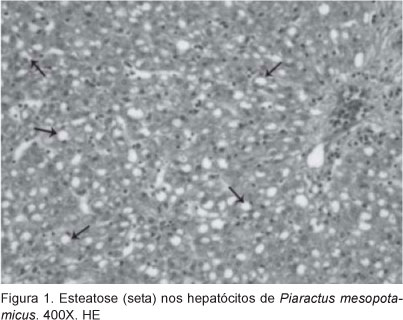
Histological analysis of kidney, spleen and liver of Piaractus mesopotamicus, Prochilodus lineatus and Pseudoplatystoma fasciatum, infected by myxosporean, caugth in Aquidauana river, MS, was studied. After necropsy, samples of liver, previous kidney and spleen were fixed in 10 % buffered formalin and processed followed histological routine methods. Sections of 5 μm were stained with hematoxylin and eosin. Myxobolusporofilus, M. colossomatis and were found in P. lineatus, in P. mesopotamicus respectively and Myxobolus spp. Were also found in all three species of fish. Myxosporideans cysts in the liver and spleen of P mesopotamicus were also related. Up to 50% of P mesopotamicus and P lineatus liver samples showed diffuse hepatodistrofy. Liver sections also showed concentric hialin structures in over 80 % of samples and esteatosis in 50% of them. In P mesopotamicus kidney, 95.23 % of them showed tissue changes consisted of 60 % with diffuse moderate nefrodistrofy and congestion of glomerular sinusoids. In P lineatus kidney, 20 % of the samples showed tissues changes. No heavy damage was observed in the fish spleen.
Histological alterations; natural environment; parasites; fishes