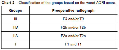
OBJECTIVE: To evaluate the value of preoperative radiographic evaluations for total knee arthroplasty (TKA) revision. METHODS: Thirty-one knees that were operated between 2006 and 2008, in a consecutive series of cases of TKA revision surgery were analyzed retrospectively. THE FOLLOWING CRITERIA WERE EVALUATED: number of wedges or structured bone grafts used for filling the bone defects; locations of the wedges and bone grafts used; and mean thickness of the polyethylene used. The AORI classification was previously established based on preoperative radiographs, using preestablished criteria. After the analysis, the knees were divided into four groups (I, IIA, IIB and III). RESULTS: The mean number of wedges or grafts used in each knee progressively increased among the groups (group I: 1.33; group IIA: 2; group IIB: 4.33; and group III: 4.83) (P = 0.0012). The commonest locations were medial in the tibia and posteromedial in the femur. There were no statistically significant differences in the thickness of the polyethylene used. CONCLUSION: The AORI classification for bone defects in the knee, based on preoperative radiographs, showed a correlation with increasing need to use wedges and/or structured grafts in TKA revisions. However, up to 46% of the knees in groups I and IIA presented bone defects of up to 5 mm that were not diagnosed by means of preoperative radiographs.
Bone Defects; Total Knee Arthroplasty Revision; Radiography