| 1. Review previous imaging studies and perform a well documented targeted ultrasonography scan. |
| 2. Evaluate whether the biopsy is appropriately indicated and its limitations (Tables 1 and 2). |
| 3. Obtain the term of free and informed consent from the patient after having explained its entire contents. |
| 4. Define the pathway to approach the lesion, as well as which of the physician's hands will be used for each function. |
| 5. Carry out the antisepsis of the transducer and prepare the materials on a portable table. |
| 6. Don the sterile gloves and couple the core biopsy needle (14G) to the device. Perform a triggering test, checking out the needle travel, as well as the triggering sound from the device. |
| 7. Aspirate the anesthetic agent (1-2% lidocaine without vasoconstrictor). |
| 8. Positioning the patient (usually in dorsal or anterior oblique decubitus). |
| 9. Perform the antisepsis over a wide area around the lesion, over which a sterile fenestrated drape should be placed. The antiseptic or sterile gel will serve as an ultrasound conductive agent. |
| 10. Sonographically identify the lesion. The palm of the hand holding the transducer and the fourth and fifth fingers exert some pressure on the breast to avoid its motion. |
| 11. Remember the access and entry point defined on item 4. Under US guidance, inject the anesthetic agent through the entire pathway up to the lesion. |
| 12. Make a 2-3 mm incision on the skin over needle entry point. |
| 13. Insert the biopsy needle through the incision, attempting to follow the same pathway of the anesthetic needle towards the lesion border. At this point, the needle is to be directed to a position parallel to the nodule. |
| 14. Tell the patient that a sample is about to be obtained, and trigger the device action. |
| 15. Cross sectionally and longitudinally slide the transducer aiming at verifying whether the needle penetrated the nodule and that no injury occurred to the chest wall. |
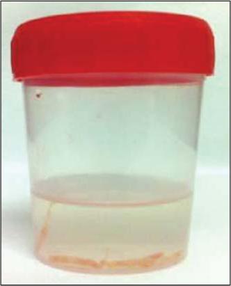
| 16. Retrieve the sample from the needle with the scalpel blade or sterile needle, placing it in the vial with formaldehyde, briefly evaluating its characteristics. |
| 17. Repeat steps 13 to 16 until a minimum of five good samples are obtained, preferably from different areas of the lesion (center, 3, 6, 9, and 12 o'clock positions). In cases of microcalcifications, at least ten samples should be collected and submitted to radiography, identifying and separating those without calcifications from the ones with calcium. |
| 18. Compress the lesion and incision areas for at least five minutes and apply local ice. |
| 19. Perform asepsis and apply compressive dressings which should be left in place for 24-48 hours. |
| 20. Instruct the patient to avoid intense physical exertion and prescribe pain-relievers and non-steroid anti-inflammatory medication, as necessary. |
| 21. Clarify doubts and schedule return as soon as the histopathological results are available. The approach should be adopted according to Table 4. |