ABSTRACT:
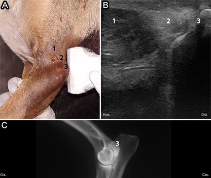
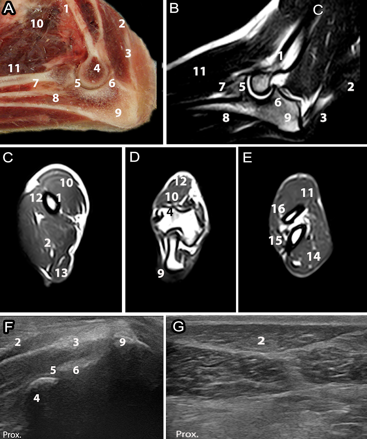
The elbow is a complex joint and has great clinical relevance in small animal medicine. Previous research in this area has been performed using radiographic and tomographic methods; however, there are limited studies on ultrasonography. The aims of this study was suggesting an evaluation protocol for elbow scan and describe the ultrasonographic anatomy of the elbow joint in dogs. Ten cross-breed dogs weighing 5-15kg underwent radiography and were selected for this ultrasonographic study. The protocol was established for the ultrasonographic description dividing the articular areas in the proximal, middle, and distal, lateral, cranial, medial, and caudal faces. The approach was performed in the longitudinal, transverse and oblique planes and the musculoskeletal structures were described according to the architecture, echogenicity and echotexture. Computed tomography and magnetic resonance imaging scans were obtained for one animal for comparison. Ultrasonography was effective in visualizing and analyzing muscles, tendons and ligaments. Bone contours and regions that have clinical significance such as the medial coronoid process and anconeus process were identified, but with limited access. Prior knowledge of the normal sonographic anatomy of the elbow joint, as well as its technical advantages and limitations will allow further studies related to the identification of musculoskeletal disorders.
INDEX TERMS:
Musculoskeletal ultrasonography; elbow joint; canine; articulation; diagnostic imaging; standardization; dogs; morphology

 Thumbnail
 Thumbnail
 Thumbnail
 Thumbnail
 Thumbnail
 Thumbnail
 Thumbnail
 Thumbnail


