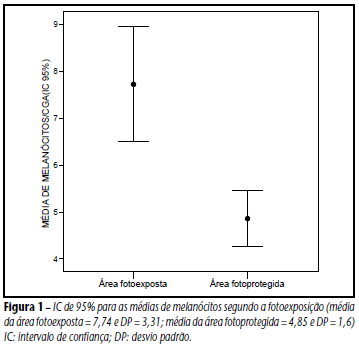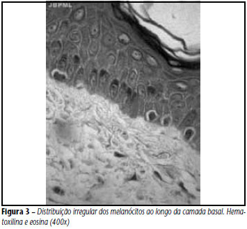
INTRODUCTION AND OBJECTIVES: It is believed that sun exposure can change the number, distribution and morphology of melanocytes in human skin, which often hinders the interpretation of skin biopsies, mainly as to diagnosis of initial melanocytic lesions and accurate assessment of resection margins. Our objective was to evaluate melanocytes in sun-exposed and non-exposed skin. METHODS: It was conducted the histological analysis of 60 skin biopsy samples resected from cadaver forearm (sun-exposed skin) and cadaver buttock (non-exposed skin) from the Death Verification Service (Serviço de Verificação de Óbitos) of Recife, state of Pernambuco. The statistical analysis was performed with SPSS Windows version 12.0. RESULTS: There was considerable variability in melanocyte density, with a higher concentration of these cells in sun-exposed areas (p < 0.001). There was also an irregular distribution of melanocytes along the epidermal basal layer, occasionally with cells arranged side by side. This confluence was identified with a higher frequency in sun-exposed areas (p = 0.035) and did not exceed more than 4 adjacent melanocytes. Cytological atypia was found in 40% of sun-exposed skin samples and was absent in non-exposed areas. Neither nests of melanocytes nor pagetoid spread was observed. CONCLUSION: There is a great variability in the density, distribution and morphology of melanocytes in human skin, mostly in sun-exposed areas. COMMENT: The presence of cytological atypia and cell confluence should not be used as isolated criteria for the definition of a neoplastic lesion
Melanocytes; Density; Morphology