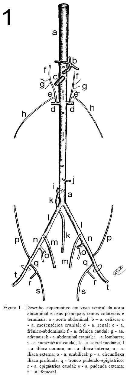
For this study it was used 30 nutria, 15 females and 15 males, with its abdominal aorta system full filled with latex 603, stained in red, and fixed in an aqueous solution of formaldehyde 20%. The abdominal aorta emitted from its dorsal surface 6 to 8 single lumbar arteries. From the renal arteries, left and right, it has been originated the phrenicoabdominal arteries, in order to irrigate part of the diaphragm and the cranial lateral abdominal walls. The abdominal aorta emitted, dorsally, the median sacral artery, cranially to its bifurcation into the common iliac arteries. These common iliac arteries, terminal branches of the aorta, originate the internal and external iliac arteries. The internal iliac artery was distributed along the pelvic cavity viscera. The external iliac artery emitted an umbilical artery and, before reaching the inguinal ring, emitted the deep circunflex iliac artery to 2/3 of the caudal lateral abdominal wall. The external iliac artery emitted the pudendoepigastric trunk, wich has originated the caudal epigastric artery, to the ventral abdominal wall, and the external pudendal artery, wich passed through the inguinal ring to irrigate the external genital. The direct parietal branches of the abdominal aorta were the lumbar arteries and the median sacral artery, while the phrenicoabdominal arteries, deep circunflex iliac and the caudal epigastric artery were indirect colateral parietal branches. The terminal branches of the abdominal aorta were the common iliac arteries with its branches, the internal and external iliac arteries.
abdominal arteries; vascularization; rodents; nutria; Myocastor coypus