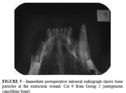
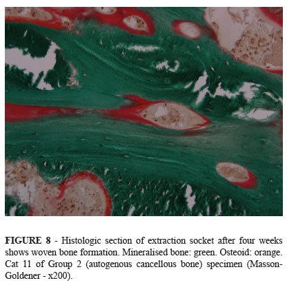
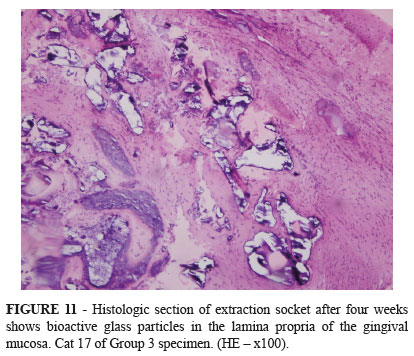
PURPOSE: To evaluate bone healing in the extraction socket of the feline maxillary canine tooth after grafting. METHODS: Eighteen adult cats were submitted to unilateral extraction of maxillary canine tooth and divided into three groups. In group 1 (n=6), control, the extraction socket was left empty. In group 2 (n=6), the extraction socket was filled with autogenous cancellous bone from the iliac crest and in group 3 (n=6), with bioactive glass particulate material. Cats were euthanized at four weeks postoperative. RESULTS: The radiographic examinations performed four weeks after surgery showed that in all groups the healing process converged to a radiopacity similar to that observed in the surrounding bones. Histological examination showed formation of woven bone within the extraction socket. The percentage of newly formed bone within the extraction socket, measured by the histometry, showed no statistically significant difference among the values of the three groups (Kruskal-Wallis'test p>0.05) (group 1: 63.96 ± 5.85, group 2: 66.84 ± 11.67, group 3: 59.28 ± 15.50). CONCLUSION: The bone regeneration observed in the extraction sockets filled with autogenous cancellous bone or bioactive glass was similar to that observed in the control sites, given an observation period of four weeks after extraction of the maxillary canine tooth.
Surgery, Oral; Bone Transplantation; Tooth Extraction; Tooth Socket; Cats