ABSTRACT
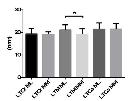
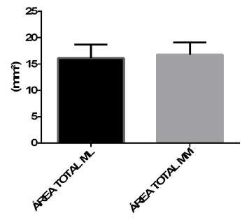
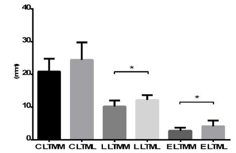
Meniscal lesions are one of the main causes of pain and lameness in horses. The objective of this study was to evaluate the morphometry of the meniscus of the femorotibial joint of horses. 48 meniscus were used from 12 animals, aged between five and 15 years and weighing between 400kg and 500kg. The measurement of the peripheral extension went from the most cranial part to the most caudal, denominated external circumference (CE). The internal margin, with the same treatment, was called internal circumference (IC). The meniscus was divided into cranial, middle and caudal thirds. The thickness was obtained at the midpoints of each third. The area of the meniscus in contact with the femoral condyles was also calculated. The medial meniscus presented a higher CE with a mean of 126.38mm, while the lateral meniscus presented a mean of 115.32mm. The lateral meniscus showed greater thickness in the middle and caudal thirds, with mean values of 16.00mm and 19.85mm respectively, against 13.75mm and 14.99mm of the medial meniscus. The results of this study showed an important relationship between the morphometric data and the clinical findings in an attempt to explain the higher incidence of lesions involving the medial meniscus.
Keywords:
horse; knee; meniscus; lameness