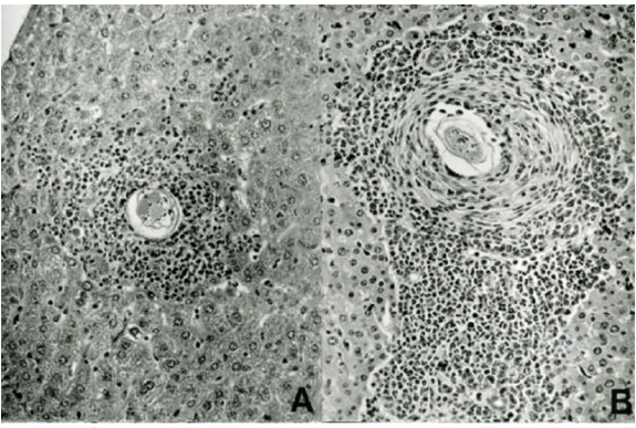
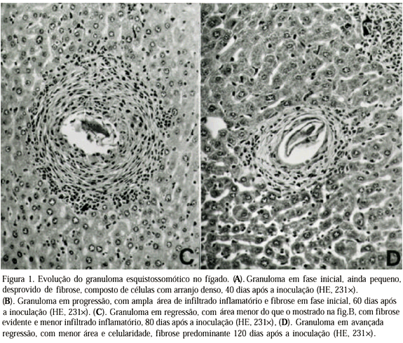
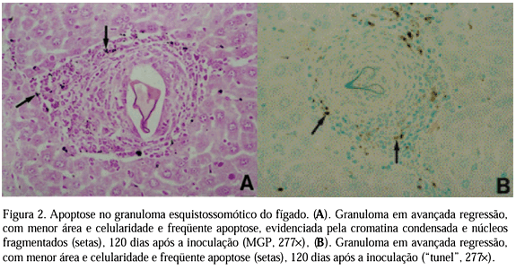
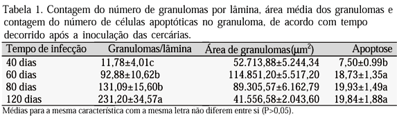
Forty two samples of liver, obtained from mice inoculated with Schistosoma mansoni cercariae, were studied morphometrically. Samples were collected from mice at 40, 60, 80 and 120 days after inoculation and processed according to routine procedures. Four m m sections were stained by hematoxiline-eosine for microscopic evaluation, and morphometry of number and area of granulomas, and by MGP for apoptotic cells quantification, considering the age of inoculation. Forty days after inoculation the animals presented less granulomas (<img src="http:/img/fbpe/abmvz/v52n6/a06img02.gif" alt="a06img02.gif (533 bytes)" align="absmiddle" > or = 11.78 ± 4.01), less apoptosis (<img src="http:/img/fbpe/abmvz/v52n6/a06img02.gif" alt="a06img02.gif (533 bytes)" align="absmiddle" > or = 7.50 ± 0.99) and smaller granulomas (<img src="http:/img/fbpe/abmvz/v52n6/a06img02.gif" alt="a06img02.gif (533 bytes)" align="absmiddle" > or = 52,713.88 ± 5,244.34mum²). At 60 days of inoculation they presented the largest granulomas (<img src="http:/img/fbpe/abmvz/v52n6/a06img02.gif" alt="a06img02.gif (533 bytes)" align="absmiddle" > or = 114,851.20 ± 5,517.20mum²), increased numbers of granulomas (<img src="http:/img/fbpe/abmvz/v52n6/a06img02.gif" alt="a06img02.gif (533 bytes)" align="absmiddle" > or = 92.88 ± 10.62) and of apoptosis (<img src="http:/img/fbpe/abmvz/v52n6/a06img02.gif" alt="a06img02.gif (533 bytes)" align="absmiddle" > or = 18.73 ± 1.35). After 80 days of inoculation the number of granulomas had increased (<img src="http:/img/fbpe/abmvz/v52n6/a06img02.gif" alt="a06img02.gif (533 bytes)" align="absmiddle" > or = 131.09 ± 15.60), presenting smaller area (<img src="http:/img/fbpe/abmvz/v52n6/a06img02.gif" alt="a06img02.gif (533 bytes)" align="absmiddle" > or = 89,305.57 ± 6,162.79mum²) and increased apoptosis (<img src="http:/img/fbpe/abmvz/v52n6/a06img02.gif" alt="a06img02.gif (533 bytes)" align="absmiddle" > or = 19.93 ±1.49). Mice at 120 days after inoculation showed the greatest quantity of granulomas (<img src="http:/img/fbpe/abmvz/v52n6/a06img02.gif" alt="a06img02.gif (533 bytes)" align="absmiddle" > or = 231.20 ± 34.57), almost the same frequency of apoptosis (<img src="http:/img/fbpe/abmvz/v52n6/a06img02.gif" alt="a06img02.gif (533 bytes)" align="absmiddle" > or = 19.84 ± 1.88), but with the smallest areas (<img src="http:/img/fbpe/abmvz/v52n6/a06img02.gif" alt="a06img02.gif (533 bytes)" align="absmiddle" > or = 41,556.58 ± 2,043.60mum²). Apoptosis was confirmed by TUNEL technique. The occurrence of apoptosis helps to explain the decrease in both cellularity and area of granulomas. It was concluded that apoptosis participates in the modulation of the granulomatous inflammatory process in Schistosomiasis as a reaction to embolization and deposition of eggs of the parasite in liver. As Schistosomiasis progresses, some immunological tolerance to Schistosoma mansoni egg antigens develops, which is detected morphologically and morphometrically by decrease in the area of the granulomas and by increase in the occurrence of apoptosis in granuloma cells.
Schistosomiasis; apoptosis; programmed cell death; modulation of inflammation; granuloma