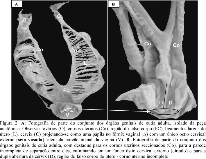
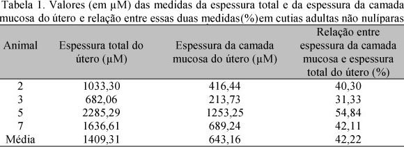
The uterine morphology was studied through ovarysalpingohysterectomy in nulliparous and non nulliparous agoutis (Dasyprocta azare). The uterus macroscopic analysis was done "in loco" and in the removed specimens. Fragments of the proximal, media and distal portions of this viscerae were collected, fixed and histologically prepared, and the samples analyzed through light microscopy and through the histomorphometry of the uterine layers. Topographycally, the uterus of this rodent is located on the sub lumbar area, caudally to the kidneys, and following the ovaries and uterine horns, getting through the pelvic entrance, where it is located dorsally to the bladder. It is characterized as a double uterus, although there is only an external cervical os. Microscopically, the uterine mucous is formed by epithelial elevations, from cylindrical to pseudostratified epithelium, which is supported by a loose connective tissue where endometrial glands covered by cylindrical epithelium can be observed, besides blood vessels. The muscle layer is subdivided in inner or submuscous, median or vascular and outer or subserous. The serous layer is composed of a connective tissue and mesothelium. In the histomorphometry analysis, the total uterine thickness and the mucous layer thickness, in average, were bigger on non nulliparous females.
Dasyprocta azare; rodent; hystricomorph; anatomy; histology